. 2002 Apr 2;99(7):4538-43.
doi: 10.1073/pnas.062621699. Epub 2002 Mar 26.

Naive T cells proliferate strongly in neonatal mice in response to self-peptide/self-MHC complexes

Affiliations

Naive T cells proliferate strongly in neonatal mice in response to self-peptide/self-MHC complexes

Armelle Le Campion et al. Proc Natl Acad Sci U S A. .

Abstract

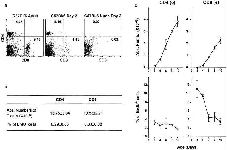
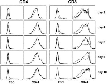
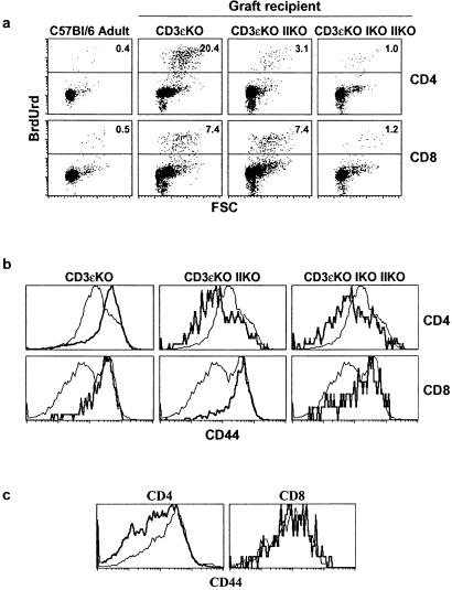
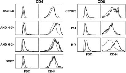
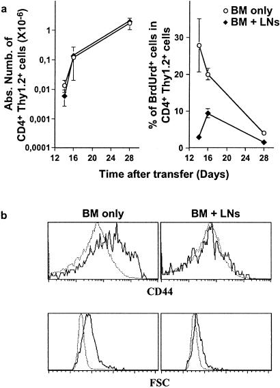
Adult naive T cells, which are at rest in normal conditions, proliferate strongly when transferred to lymphopenic hosts. In neonates, the first mature thymocytes to migrate to the periphery reach a compartment devoid of preexisting T cells. We have extensively analyzed the proliferation rate and phenotype of peripheral T cells from normal C57BL/6 and T cell antigen receptor transgenic mice as a function of age. We show that, like adult naive T cells transferred to lymphopenic mice, neonatal naive T cells proliferate strongly. By using bone-marrow transfer and thymic-graft models, we demonstrate that the proliferation of the first thymic emigrants reaching the periphery requires T cell antigen receptor-self-peptide/self-MHC interactions and is regulated by the size of the peripheral T cell pool.


Figures

Figure 1
Figure 1
Peripheral T cells proliferate strongly in neonates. The absolute number and proliferation rate of peripheral T cells from normal C57BL/6 mice were studied from 2 to 10 days after birth and in adult control mice. To detect and characterize DNA-synthesizing cells, BrdUrd was injected i.p. twice, at a 30-min interval. Spleens were taken 30 min after the second injection, cell-counted, and stained for CD4, CD8, TCR surface expression, and BrdUrd incorporation. (a) CD4/CD8 fluorescence dot-plots of adult and day-2 normal and nude C57BL/6 mice. (b) Absolute numbers of splenic CD4+ and CD8+ TCRhi cells and percentages of BrdUrd+ cells among these subsets in adult mice. (c) Absolute numbers of splenic CD4+ and CD8+ TCRhi cells and percentages of BrdUrd+ cells among these subsets as a function of age.
Figure 2
Figure 2
CD44 expression and size of neonatal spleen CD4+and CD8+ T cells. From 2 to 10 days after birth, spleens from normal C57BL/6 mice were taken and stained for CD4, CD8, TCR, and CD44 surface expression. Forward scatter (FSC) and CD44 fluorescence histograms of CD4+ and CD8+ TCRhi cells from 2- to 10-day-old mice (thick lines) in comparison with FSC and CD44 fluorescence histograms of adult mouse CD4+ and CD8+ T cells (thin lines).
Figure 3
Figure 3
Peripheral T cells proliferate in neonates in response to TCR/MHC interactions. Thymuses from 2- to 3-day-old C57BL/6 mice were engrafted under the kidney capsule of normal CD3ɛ−/− mice and CD3ɛ−/− mice lacking MHC class II molecules or MHC classes I and II molecules. One week after grafting, spleens and thymuses were removed, cell-counted, and stained for CD4, CD8, TCR, and CD44 expression or BrdUrd incorporation. (a) FSC/BrdUrd fluorescence dot-plots of both spleen CD4+ and CD8+ TCRhi cells. (b) CD44 fluorescence histograms of graft-derived CD4+ and CD8+ TCRhi spleen cells (thick lines) in comparison with CD44 expression on adult CD4+ and CD8+ spleen T cells (thin lines). (c) CD44 fluorescence histograms of graft-derived CD4+ and CD8+ TCRhi thymocytes (thick lines) in comparison with CD44 expression on adult CD4+ and CD8+ TCRhi thymocytes (thin lines).
Figure 4
Figure 4
Peripheral naive T cells proliferate strongly in neonates. Two days after birth, spleens from different mouse strains were taken and stained for CD4, CD8, TCR, and CD44 surface expression. FSC and CD44 fluorescence histograms of CD4+ TCRhi spleen cells from 2-day-old mice of different strains (thick lines). FSC and CD44 fluorescence histograms of CD4+ TCRhi spleen cells from the corresponding adult mice are shown as controls (thin lines). FSC and CD44 fluorescence histograms of CD8+ TCRhi spleen cells from 2-day-old mice of different strains (thick lines). FSC and CD44 fluorescence histograms of CD8+ TCRhi spleen cells from the corresponding adult mice are shown as controls (thin lines).
Figure 5
Figure 5
The extent of thymic emigrant proliferation depends on the size of the peripheral T cell pool. H-2b CD3ɛ-deficient mice were irradiated and injected with CD4- and CD8-depleted BM cells from H-2b AND TCR transgenic mice (Thy 1.2). When indicated, 20 × 106 lymph node T cells from C57BL/6 Ba mice (Thy 1.1) were coinjected. Spleens of these mice were recovered 14–28 days after BM transfer. (a) Absolute numbers of splenic CD4+ Thy 1.2+ cells and percentages of BrdUrd+ cells among this subset as a function of time after transfer. (b) FSC and CD44 fluorescence histograms of day 16 after transfer of CD4+ Thy 1.2+ splenic cells (plain lines) in comparison with FSC and CD44 fluorescence histogram of CD4+ splenic cells from H-2b AND TCR transgenic adult mice (dotted line).

Similar articles

Cited by

References

    1. Tanchot C, Rosado M M, Agenes F, Freitas A A, Rocha B. Semin Immunol. 1997;9:331–337. - PubMed
    1. Tanchot C, Rocha B. Immunol Today. 1998;19:575–580. - PubMed
    1. Tanchot C, Rocha B. J Exp Med. 1997;186:1099–1106. - PMC - PubMed
    1. Von Boehmer H, Hafen K. J Exp Med. 1993;177:891–896. - PMC - PubMed
    1. Tough D F, Sprent J. J Exp Med. 1994;179:1127–1135. - PMC - PubMed

Publication types

Substances