. 2002 Apr 2;99(7):4331-6.
doi: 10.1073/pnas.072088099. Epub 2002 Mar 26.

The procaspase-8 isoform, procaspase-8L, recruited to the BAP31 complex at the endoplasmic reticulum

Affiliations

The procaspase-8 isoform, procaspase-8L, recruited to the BAP31 complex at the endoplasmic reticulum

David G Breckenridge et al. Proc Natl Acad Sci U S A. .

Abstract

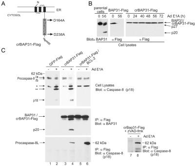
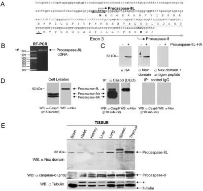
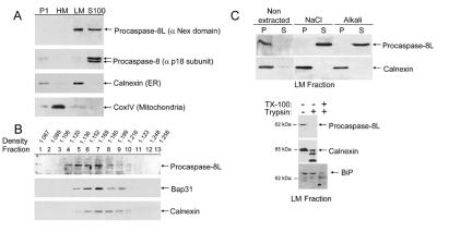
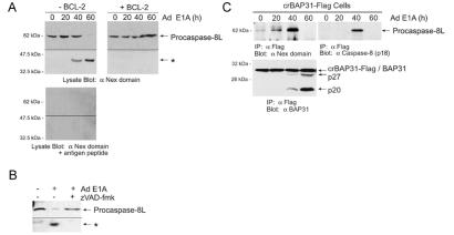
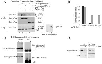
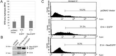
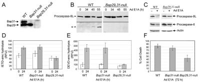
BAP31 is an integral protein of the endoplasmic reticulum membrane and a substrate of caspase-8. Here, we describe the procaspase-8 isoform, procaspase-8L, which is ubiquitously expressed and selectively recruited to the BAP31 complex in response to apoptotic signaling by E1A. Procaspase-8L is characterized by the N-terminal extension (Nex) domain, which extends procaspase-8/a at the N terminus and is required for selective association of procaspase-8L with the BAP31 complex. Gene deletion identified BAP31 and related BAP29 as required for processing of procaspase-8L in response to E1A, by a FADD-independent mechanism that was blocked by BCL-2. Further, Bap29,31 deletion, as well as a Nex-domain dominant-negative mutant, curtailed the activation of downstream caspases (IETDase and DEVDase) and cell death in response to E1A. Preferential recruitment of procaspase-8L by the BAP31 complex at the endoplasmic reticulum suggests an additional pathway for regulating initiator caspase-8 during apoptosis.


Figures

Figure 1
Figure 1
Procaspase-8L is recruited to the BAP31 complex during E1A-induced apoptosis. (A) Schematic of crBAP31-Flag in the ER. Asp-164 and Asp-238 were mutated to Ala, and a Flag epitope was inserted directly in front of the KKEE ER retrieval sequence. *, Caspase recognition Asp residues. (B) crBAP31-Flag is not cleaved during E1A-induced apoptosis. Parental KB cells or KB cells stably expressing wt BAP31-Flag or crBAP31-Flag (at levels slightly higher than that of endogenous BAP31) were treated with adenovirus type 5 (Ad5) dl520E1B (expressing only 12 S E1A and no E1B products) for the indicated times and endogenous BA, 31, BAP31-Flag, or crBAP31-Flag cleavage was analyzed by SDS/PAGE and immunoblotting with anti-BAP31 or anti-Flag antibodies. (C) A ≈62-kDa form of procaspase-8, designated procaspase-8L, is recruited to the BAP31 complex in response to E1A signaling. KB cells stably expressing GFP-Flag, crBAP31-Flag, or crBAP31-Flag and BCL-2 were mock infected or infected (E1A) with Ad5 dl520E1B for 40 h and immunoprecipitation (IP) was conducted with anti-Flag M2 gel followed by SDS/PAGE and Western blotting with anti-BAP31 (Middle) or anti-caspase-8 antibody specific for the p18 subunit (Bottom). Where indicated, cells were cultured in the presence of zVAD-fmk (50 μm). Immunoblots of the corresponding total cell lysate (5% of the input used for immunoprecipitation) are also shown (Top). The positions of the conventional procaspase-8 isoforms, designated/a and/b, their processing intermediates (asterisks), the caspase-8 catalytic subunit (p18), procaspase-8L, crBAP31-Flag, endogenous BAP31, and its p20 cleavage product, are shown.
Figure 2
Figure 2
Characterization and cloning of procaspase-8L. (A) Deduced amino acid sequence of the predicted N-terminal extension (Nex) domain of procaspase-8L. The predicted procaspase-8L upstream start codon and Kozak translation initiation consensus sequence are shown in bold. The positions of exon 3 and the canonical procaspase-8/a and -8/b start site are indicated. The sequence of the peptide used for the procaspase-8L Nex domain antibody production is underlined in black. (B) PCR of first-strand cDNA derived from KB cell mRNA by using primers specific for the 5′ end of the Nex domain and the 3′ end of the canonical caspase-8 p10 subunit demonstrate that the Nex domain of procaspase-8L is connected to the entire procaspase-8/a ORF. The reverse transcription (RT)-PCR product is indicated (arrow). (C) Affinity-purified rabbit polyclonal antibodies raised to the Nex domain of procaspase-8L detect an endogenous protein of ≈62 kDa in human H1299 lung carcinoma cells (arrow, Center) and ectopic expressed procaspase-8L-HA. Cells were transfected with vector (−) or with vector expressing HA-tagged procaspase-8L (+) in the presence of 50 μM zVAD-fmk and, after 24 h, total cell lysates were analyzed by Western blotting with the indicated antibodies, in the presence or absence of 10 μM immunizing peptide. (D) Cell lysates were subjected to immunoprecipitation with an anti-caspase-8 monoclonal antibody recognizing the prodomain (10). Lysates and precipitates were analyzed by blotting with anti-caspase-8 p18 subunit-specific antibody or anti-Nex domain antibody. (E) Tissue distribution of procaspase-8L. The indicated murine tissue lysates were analyzed by immuno blotting as in D. The blot was reprobed with antitubulin monoclonal antibody for loading control. *, Cross-reacting product.
Figure 3
Figure 3
Subcellular distribution of procaspase-8L. (A) KB cells were homogenized in isotonic buffer and the P1 nuclear (500 × g pellet), heavy membrane (HM) (9,000 × g pellet), LM (100,000 × g pellet), and cytosolic S-100 fractions were separated by differential centrifugation. The fractions were analyzed for the presence of procaspase-8L, procaspase-8, the ER marker, calnexin, and the mitochondrial marker, cytochrome c oxidase subunit IV (CoxIV), by immunoblotting with the respective antibodies. (B) Sucrose density gradient fractions of rodent liver membranes were analyzed for the presence of procaspase-8L, BAP31, and calnexin as in A. (C) Procaspase-8L is peripherally associated with the cytosolic face of the light membranes. (Upper) The LM fraction from KB cells was subjected to extraction with 0.5M NaCl or alkali (0.1 M NaCO3, pH 11.5) and, after centrifugation, the membrane pellets (P; lanes 1 and 3) and supernatants (lanes 2 and 4) were analyzed as in A. (Lower) The LM fraction was incubated with (+) or without (−) trypsin and the integrity of procaspase-8L, calnexin (ER transmembrane protein), and BiP (ER luminal protein) assessed by immunoblotting. TritonX-100 was added to 1% in one reaction to demonstrate that BiP was trypsin sensitive following solubilization of the LM microsomal fraction.
Figure 4
Figure 4
Endogenous procaspase-8L is recruited to the BAP31 complex and cleaved during E1A-induced apoptosis. (A) Cleavage of procaspase-8L is blocked by BCL-2. Parental KB cells (−BCL-2) or KB cells stably expressing BCL-2 (+BCL-2) were infected with Ad5 dl520E1B for the indicated times and Western blots were developed with anti-Nex antibody, in the absence (Upper) or presence (Lower) of the Nex immunizing peptide (10 μM). (B) Cleavage of procaspase-8L is inhibited by z-VAD-fmk. Cells were treated as in A in the absence or presence of 50 μM zVAD-fmk. (C) Recruitment of procaspase-8L to the BAP31 complex. KB cells stably expressing crBAP31-Flag were treated as in A and the BAP31 complex was immunoprecipitated (α Flag IP) at the indicated times postinfection. Immuno blots of the precipitates were developed with anti-Nex antibody (Upper Left), then stripped and reprobed with anti-caspase-8 (p18) antibody (Upper Right) or anti-BAP31 antibody (Lower). The anti-Nex domain and anti-caspase-8 (p18) antibodies detected an identical 62-kDa protein. The p20 and p27 cleavage products of endogenous BAP31 (that heterodimerize with crBAP31-Flag) are indicated. A representative experiment is shown.
Figure 5
Figure 5
Procaspase-8L selectively associates with the BAP31 complex. (A) Anti-Flag immunoprecipitates from H1299 cells cotransfected with the indicated expression constructs in the presence of 50 μM zVAD-fmk were analyzed by SDS/PAGE and Western blotting with anti-HA or anti-Nex domain antibodies. Note that only procaspase-8L-HA immunoprecipitated with BAP31-Flag. *, A nonspecific protein detected by anti-HA antibody in lysates. (B) Procaspase-8L induces loss of cell viability when ectopically expressed. H1299 cells were transfected with a luciferase expression construct and pcDNA3 vector expressing either procaspase-8/a-HA or -8L-HA, in the absence or presence of 50 μM zVAD-fmk, and luciferase activity was measured 24 h later. (C) Procaspase-8L is not detected in the Fas DISC. The DISC was immunoprecipitated following stimulation of H9 lymphocytes with anti-Apo-1/Fas (19) and the presence of procaspase-8L and procaspase-8 in the precipitates was assessed by immunoblotting as in Fig. 4C. IgG, Ig heavy chain. (D) Wt or Fadd-deficient mouse embryo fibroblasts (20) were infected with Ad5 dl520E1B for 50 h and procaspase-8L cleavage was analyzed as in Fig. 4A.
Figure 6
Figure 6
E1A-induced caspase-8 activity and apoptosis is inhibited by a procaspase-8L DN mutant. (A) KB cells were transiently cotransfected with plasmids encoding 12S E1A and Nex-Flag-EGFP or 12S E1A and Flag-EGFP and 36 h post transfection equivalent amounts of cell lysate were tested for their ability to hydrolyze the caspase-8-preferred substrate IETD-amc. Shown is the mean of four independent experiments. (B) Cell lysates from A were analyzed by SDS/PAGE and immunoblotting with antibodies against E1A or Flag. (C) As in A, except that cells were collected, stained with Annexin V, and analyzed by flow cytometry. Transfection efficiency was estimated to be 20–30% by analyzing EGFP-positive cells by immunofluorescence (not shown).
Figure 7
Figure 7
Procaspase-8L processing is inhibited in Bap29,31-null cells. (A) Loss of Bap29 and Bap31 expression was confirmed by immunoblotting with anti-BAP31 and anti-Bap29 antibodies. (B) E1A-induced procaspase-8L cleavage is inhibited in Bap29,31-null ES cells. ES cells were differentiated into epithelial- and fibroblast-like cells as described in Materials and Methods, and infected with Ad5 dl520E1B for the indicated times. Procaspase-8L cleavage was analyzed as in Fig. 3A. (C) As in B, procaspase-8L and procaspase-8/a and -8/b cleavage was analyzed with anti-Nex and anti-caspase-8 (p18) antibodies. (D) Decreased caspase-8 activity in Bap29,31-null cells. Aliquots of lysate from wild-type and Bap29,31-null cells (containing equivalent protein concentrations) stimulated with E1A for 24h (time of maximum caspase activity) were tested for their ability to hydrolyze the preferred caspase-8 substrate IETD-amc. Shown is the average of three independent experiments. RFU, relative fluorescence units. (E) Decreased DEVD-ase activity in Bap29,31-null cells. As in C, except that lysates were tested for their ability to hydrolyze the caspase-3 preferred substrate DEVD-amc. (F) Cell death was measured by trypan blue exclusion 72 h post infection. Shown is a representative of four independent experiments.

References

    1. Budihardjo I, Oliver H, Lutter M, Luo X, Wang X. Annu Rev Cell Dev Biol. 1999;15:269–290. - PubMed
    1. Earnshaw W C, Martins L M, Kaufmann S H. Annu Rev Biochem. 1999;68:383–424. - PubMed
    1. Korsmeyer S J, Wei M C, Saito M, Weiler S, Oh K J, Schlesinger P H. Cell Death Differ. 2000;12:1166–1173. - PubMed
    1. Kim K M, Adachi T, Nielsen P J, Terashima M, Lamers M C, Kohler G, Reth M. EMBO J. 1994;13:3793–3800. - PMC - PubMed
    1. Mosser J, Sarde C O, Vicaire S, Yates J R, Mandel J L. Genomics. 1994;22:469–471. - PubMed

Publication types

MeSH terms

LinkOut - more resources