

Feline inflammatory bowel disease: a review
- PMID: 11919030
- PMCID: PMC10832802
- DOI: 10.1016/S1098-612X(99)90204-8
Feline inflammatory bowel disease: a review
Abstract
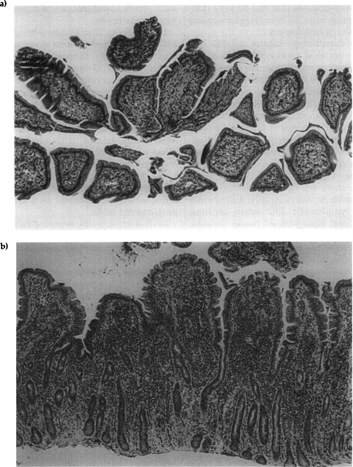
Inflammatory bowel disease (IBD), while a popular diagnosis, may not occur as commonly as it is diagnosed. It is a diagnosis of exclusion, meaning that it is important to eliminate diseases that mimick it. Dietary intolerance or allergy in particular, can have the same clinical and histologic appearance as IBD. Likewise, well-differentiated alimentary lymphosarcoma can also be confused with it. Intestinal biopsies are useful, but must be taken carefully and then evaluated by someone with interest and expertise in alimentary tract pathology. Therefore, it behoves the clinician to carefully consider the diagnosis instead of starting multiple drug therapy in a cavalier fashion. Well constructed dietary therapy can often be beneficial for both dietary problems and IBD.
Figures
References
-
- Dennis JS, Kruger JM, Mullaney TP. (1992) Lymphocytic/plasmacytic gastroenteritis in cats: 14 cases (1985–1990). Journal of the American Veterinary Medicine Association 200, 1712–1718. - PubMed
-
- Dennis JS, Kruger JM, Mullaney TP. (1993) Lymphocytic/plasmacytic colitis in cats: 14 cases (1985–1990). Journal of the American Veterinary Medicine Association 202, 313–317. - PubMed
-
- Ewing GO. (1972) Feline ulcerative colitis: A case report. Journal of the American Animal Hospital Association 8, 64–65.
-
- Ghermai AK. (1989) Chronisch-entzundliche darmerkrankungen der katze. Tierarztl Prax 17, 195–199. - PubMed
Publication types
MeSH terms
Substances
LinkOut - more resources
Full Text Sources
