. 2002 Apr 8;445(3):227-37.
doi: 10.1002/cne.10166.

Vesicular gamma-aminobutyric acid transporter expression in amacrine and horizontal cells

Affiliations

Vesicular gamma-aminobutyric acid transporter expression in amacrine and horizontal cells

Juan G Cueva et al. J Comp Neurol. .

Abstract

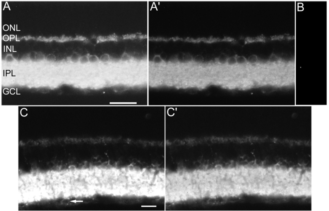
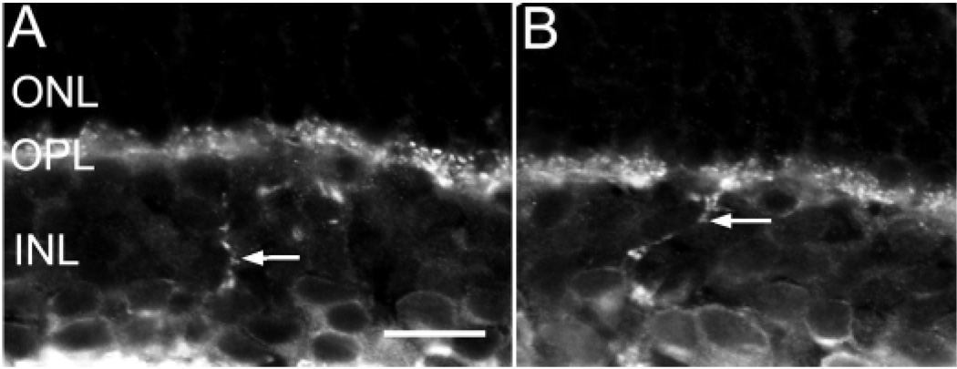
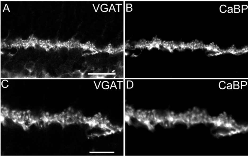
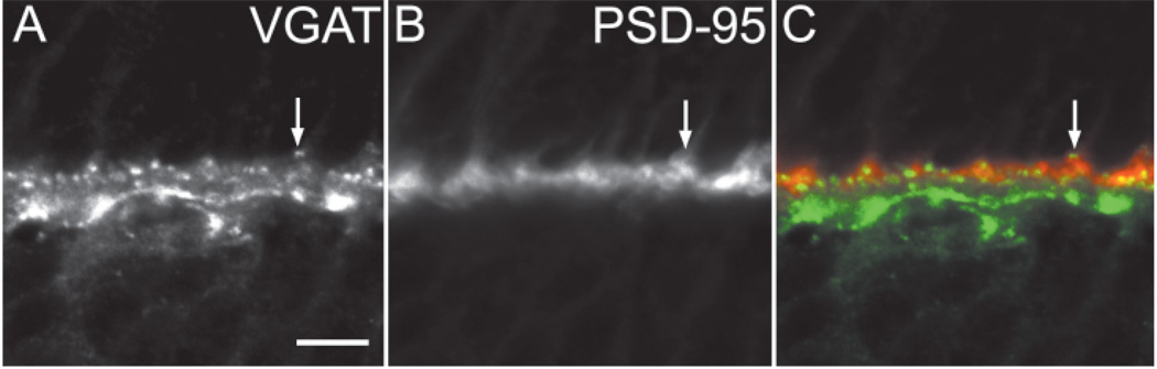
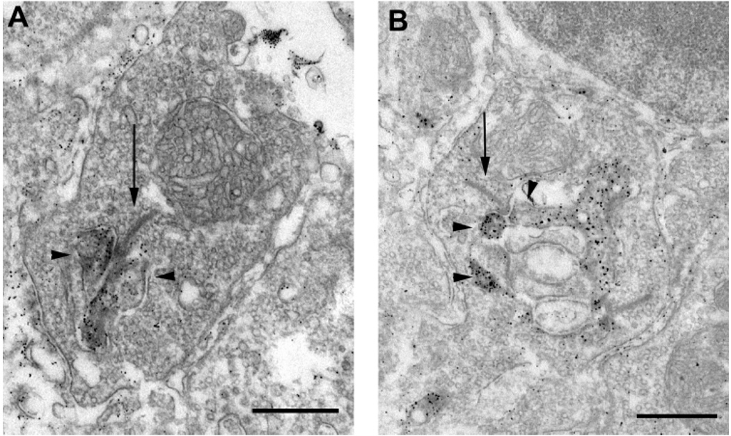
The vesicular gamma-aminobutyric acid (GABA) transporter (VGAT), which transports the inhibitory amino acid transmitters GABA and glycine, is localized to synaptic vesicles in axon terminals. The localization of VGAT immunoreactivity to mouse and rat retina was evaluated with light and electron microscopy by using well-characterized VGAT antibodies. Specific VGAT immunoreactivity was localized to numerous varicose processes in all laminae of the inner plexiform layer (IPL) and to the outer plexiform layer (OPL). Amacrine cell somata characterized by weak VGAT immunoreactivity in the cytoplasm were located in the ganglion cell layer and proximal inner nuclear layer (INL) adjacent to the IPL. In rat retina, VGAT-immunoreactive cell bodies also contained GABA, glycine, or parvalbumin (PV) immunoreactivity, suggesting vesicular uptake of GABA or glycine by these cells. A few varicose VGAT-immunoreactive processes entered the OPL from the IPL. VGAT immunoreactivity in the OPL was predominantly localized to horizontal cell processes. VGAT and calcium binding protein-28K immunoreactivities (CaBP; a marker for horizontal cells) were colocalized in processes and terminals distributed to the OPL. Furthermore, VGAT immunoreactivity overlapped or was immediately adjacent to postsynaptic density-95 (PSD-95) immunoreactivity, which is prominent in photoreceptor terminals. Preembedding immunoelectron microscopy of mouse and rat retinae showed that VGAT immunoreactivity was localized to horizontal cell processes and their terminals. Immunoreactivity was distributed throughout the cytoplasm of the horizontal cell processes. Taken together, these findings demonstrate VGAT immunoreactivity in both amacrine and horizontal cell processes, suggesting these cells contain vesicles that accumulate GABA and glycine, possibly for vesicular release.


Figures

Fig. 1
Fig. 1
A–C′: Vesicular γ-aminobutyric acid transporter (VGAT) immunoreactivity in rat and mouse retina. A: Rat retina: VGAT immunoreactivity is present in cell somata in the INL. Immunoreactivity is also present in all laminae of the IPL and the OPL. Brightness and contrast were increased to illustrate the detail of VGAT immunoreactivity in the INL and OPL. A′: The brightness and contrast were reduced in the same rat retinal section in A to illustrate the VGAT immunoreactivity pattern in all lamina of the IPL. B: Control section; the VGAT antibody was preadsorbed with the antigen, resulting in the loss of specific immunostaining. C: Mouse retina: VGAT immunoreactivity is present with the same distribution as in rat. Brightness and contrast were maximized to show the VGAT-immunoreactivity pattern in the INL and OPL. The arrow indicates a cell body in the GCL with weak VGAT immunoreactivity. C′: The brightness and contrast were minimized in the same rat retinal section in C to illustrate the VGAT-immunoreactivity pattern in all laminae of the IPL. GCL, ganglion cell layer; IPL, inner plexiform layer; INL, inner nuclear layer; OPL, outer plexiform layer; ONL, outer nuclear layer. Scale bars = 20 µm in A (applies to A–B), in C (applies to C,C).
Fig. 2
Fig. 2
A–D: Vesicular γ-aminobutyric acid (GABA) transporter (VGAT) immunoreactivity is present in GABA- and glycine-containing amacrine cells. Confocal fluorescent photomicrographs of vertical sections of the rat retina double labeled with antibodies for VGAT and GABA (A,B), VGAT and glycine (C,D), and VGAT and PV (E,F). VGAT (A) and GABA (B) immunoreactivity are colocalized in somata located in the proximal inner nuclear layer (INL) and ganglion cell layer (GCL). VGAT (C) and glycine (D) immunoreactivity are colocalized in two layers of amacrine cells in the proximal INL. VGAT (E) and PV (F) immunoreactivity are colocalized in glycine-containing AII amacrine cell somata and processes in the inner plexiform layer. Scale bars = 20 µm.
Fig. 3
Fig. 3
A,B: Vesicular γ-aminobutyric acid transporter (VGAT) immunoreactivity is present in amacrine cell somata and interplexiform processes (arrows) in the INL. INL, inner plexiform layer; OPL, outer plexiform layer; ONL, outer nuclear layer. Scale bar = 20 µm.
Fig. 4
Fig. 4
A–D: Vesicular γ-aminobutyric acid transporter (VGAT) immunoreactivity in the outer retina. Confocal fluorescent photomicrographs of a vertical section through a rat retina double labeled with antibodies to VGAT and calcium binding protein-28K (CaBP). A: Punctate VGAT immunoreactivity is present in the outer plexiform layer (OPL). B: CaBP immunoreactivity has a punctate staining pattern in the OPL virtually identical to the VGAT immunoreactivity pattern. C,D: Higher magnification of VGAT (C) and CaBP (D) immunoreactivity colocalization in the OPL. Scale bars = 20 µm in A (applies to A,B), 10 µm in C (applies to C,D).
Fig. 5
Fig. 5
A–C: Vesicular γ-aminobutyric acid transporter (VGAT) and postsynaptic density-95 (PSD-95) immunoreactivity in the outer retina. VGAT immunoreactivity is found immediately adjacent to or within PSD-95 immunoreactivity. There is overlap between the distal VGAT puncta in horizontal cells and PSD-95 immunoreactivity in photoreceptor terminals (C). The arrows point to a region of the outer plexiform layer with overlap of VGAT and PSD-95 immunoreactivity. Scale bar = 10 µm in A (applies to A–C).
Fig. 6
Fig. 6
A,B: Vesicular γ-aminobutyric acid transporter (VGAT) immunoreactivity in horizontal cell tips. Electron photomicrographs illustrate VGAT immunoreactivity in terminals of horizontal cells in the OPL of the mouse (A) and rat (B) retina. VGAT immunoreactivity is concentrated in the cytoplasm of both horizontal cell processes that invaginate rod spherules. Arrows indicate synaptic ribbons. Arrow-heads indicate horizontal cells. Scale bars = 0.5 µm in A,B.

References

    1. Agardh E, Bruun A, Ehinger B, Ekstrom P, van Veen T, Wu JY. Gamma-aminobutyric acid- and glutamic acid decarboxylase-immunoreactive neurons in the retina of different vertebrates. J Comp Neurol. 1987;258:622–630. - PubMed
    1. Attwell D, Barbour B, Szatkowski M. Nonvesicular release of neurotransmitter. Neuron. 1993;11:401–407. - PubMed
    1. Brandon C. Retinal GABA neurons: localization in vertebrate species using an antiserum to rabbit brain glutamate decarboxylase. Brain Res. 1985;344:286–295. - PubMed
    1. Brandstätter JH, Lohrke S, Morgans CW, Wässle H. Distributions of two homologous synaptic vesicle proteins, synaptoporin and synapto-physin, in the mammalian retina. J Comp Neurol. 1996;370:1–10. - PubMed
    1. Brandstätter JH, Fletcher EL, Garner CC, Gundelfinger ED, Wässle H. Differential expression of the presynaptic cytomatrix protein bassoon among ribbon synapses in the mammalian retina. Eur J Neurosci. 1999;11:3683–3693. - PubMed

Publication types

MeSH terms