. 2002 Apr 1;22(7):2469-77.
doi: 10.1523/JNEUROSCI.22-07-02469.2002.

Specificity of glomerular targeting by olfactory sensory axons

Affiliations

Specificity of glomerular targeting by olfactory sensory axons

Helen B Treloar et al. J Neurosci. .

Abstract

Axons from olfactory sensory neurons (OSNs) expressing a specific odorant receptor (OR) project to specific subsets of glomeruli in the olfactory bulb (for review, see Mombaerts, 1999, 2001). The aim of this study was to examine the trajectories that subsets of axons from OSNs expressing the same OR follow within the olfactory nerve and olfactory nerve layer (ONL) of adult mice. Using confocal microscopy, we generated serial reconstructions of axons from M72-IRES-tauGFP-expressing OSNs as they coursed within the ONL and into glomeruli. GFP-expressing axons were loosely aggregated in the outer ONL; however, as they entered the inner ONL, the majority fasciculated with other GFP-expressing axons before entering the glomerular neuropil. Although the vast majority of axons entered the glomerulus from the directly apposed ONL, some followed tortuous courses through and/or around adjacent glomeruli before terminating in the target glomerulus. Similar observations were made on subpopulations of axons in M71-IRES-tauGFP and P2-IRES-tauGFP mice. Ultrastructural analyses of labeled M72 glomeruli showed no evidence of axodendritic synapses other than those with GFP-labeled axon terminals. These data are consistent with the notion that OSN axons are highly precise in targeting glomeruli and that glomeruli, in turn, are highly homogeneous with regard to the OR expressed by the innervating OSNs. Because some single axons could follow idiosyncratic trajectories to the target glomerulus, it appears that stable homotypic fasciculation is not a prerequisite for correct targeting.


Figures

Fig. 1.
Fig. 1.
Trajectories of M72-IRES-tauGFP axons in the ONL of the adult OB. A, Fifteen serial 2 μm optical sections through part of a medial M72-IRES-tauGFP glomerulus that have been projected into a single image. Axons can been seen coursing individually in the outer ONL (ONLo;arrow) but appear to be mostly fasciculated in the inner ONL (ONLi; white arrowheads). Some axons follow torturous paths to the glomerulus through the glomerular layer (open arrowheads). B–I, To resolve individual fibers, we examined confocal images of single optical planes from the projected stack (boxed region inA). Individual fibers can be seen in the outer ONL (arrows; B–I), whereas the majority of axons fasciculate after entering the inner ONL (white arrowheads; B–I). Note that, in all panels, the outer ONL lies to theright of the inner ONL. Scale bars: A, 50 μm; B–I, 20 μm.
Fig. 2.
Fig. 2.
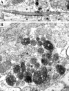
Ultrastructural immunolocalization of GFP in the ONL. A, A single GFP-expressing axon (white asterisk) coursing in the outer nerve layer among unlabeled OSN axons (e.g., black asterisks). Note the localization to microtubules, reflective of the tauGFP construct used to generate these mice. B, Within the inner ONL, labeled axons (e.g.,white asterisks) are generally found in bundles, tightly apposed to other GFP-expressing axons. Some unlabeled OSN axons are also present (e.g., black asterisks). Scale bars, 0.5 μm
Fig. 3.
Fig. 3.
Projected Z-series through medial (A, a, B,b) and lateral (C, c,D, d) M72-IRES-tauGFP glomeruli. The projections of GFP-expressing axons were followed through three serial 50 μm vibratome sections (e.g., A, A′,A") spanning the entire glomerular volume. We present lower-magnification (A–A", B–B",C–C", D–D") images so that axons distant from the glomerular target can be viewed, as well as higher-magnification images (a–a",b–b", c–c", d–d") so that single axons around glomeruli can be resolved. In all glomeruli examined, a small number of axons are observed that enter the glomerular layer from regions other than the apposed ONL (seearrowheads). These atypically projecting axons were identified in all glomeruli examined, and, although many followed tortuous paths, they all appear to target the appropriate glomerulus. Note the variation in glomerular shape and volume between the individual glomeruli. Scale bars, 50 μm.
Fig. 4.
Fig. 4.
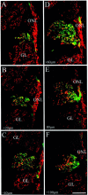
Projected Z-series through M71-IRES-tauGFP glomeruli (A–C) and P2-IRES-tauGFP glomeruli (D–F). Atypically projecting axons (seearrowheads) were also identified in mice that expressed tauGFP under the control of either the closely related M71 OR or the unrelated P2 OR. Scale bar, 50 μm.
Fig. 5.
Fig. 5.
Serial 20 μm cryostat sections (A–F) through a lateral M72-IRES-tauGFP glomerulus stained with an antibody to OMP (red). OSN axons expressing GFP (green) and following atypical paths also express OMP (e.g., as seen in D). Note that each of these images are single optical planes of ∼1 μm. Scale bars, 50 μm.
Fig. 6.
Fig. 6.
Serial 20 μm cryostat sections through a lateral M72-IRES-tauGFP glomerulus stained with an antibody to OMP. Of the six glomeruli analyzed in this way (3 medial and 3 lateral), only this glomerulus displayed clear instances of GFP-negative axons within the M72 glomerulus (see arrowheads). These axons were clearly within the glomerulus (see arrowheads inC); however, they occupied only a small portion of the entire glomerular volume. Note that these images are single optical planes of ∼1 μm. Scale bars, 50 μm.
Fig. 7.
Fig. 7.
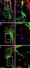
Examples of GFP-expressing axons projecting through glomeruli adjacent to an M72-IRES-tauGFP glomerulus.A, An example of a fasciculated bundle of GFP-expressing axons passing through a glomerulus en route to the target glomerulus.B, An example of an individual GFP-expressing axon within a glomerulus adjacent to the target glomerulus.C, An example of a fasciculated bundle of GFP-expressing axons with branch points within an adjacent glomerulus.a, b, and c are enlargements of boxed regions of A,B, and C, respectively. Note that these images are single optical planes of ∼1 μm. Scale bars:A–C, 50 μm; a–c, 25 μm.
Fig. 8.
Fig. 8.
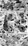
Synaptic profiles of GFP-expressing axons within M72 glomeruli. A–C, Examples of GFP-stained axons (i.e., electron dense) making Gray type 1 synapses with the electron lucent dendrites of mitral–tufted cells. The polarity of the synapses is indicted with an arrow. D, An example of unstained axons from an adjacent glomerulus. Note the difference in the electron density of the stained terminals in A–Cversus the unstained terminals in D. De, Mitral–tufted cell dendrite; Ax, axon; →, Gray type 1 synapse. Scale bars, 0.5 μm

References

    1. Berod A, Hartman BK, Pujol JF. Importance of fixation in immunohistochemistry: use of formaldehyde solutions at variable pH for the localization of tyrosine hydroxylase. J Histochem Cytochem. 1981;29:844–850. - PubMed
    1. Conzelmann S, Levai O, Bode B, Eisel U, Raming K, Breer H, Strotmann J. A novel brain receptor is expressed in a distinct population of olfactory sensory neurons. Eur J Neurosci. 2000;12:3926–3934. - PubMed
    1. Firestein S. How the olfactory system makes sense of scents. Nature. 2001;413:211–218. - PubMed
    1. Gad JM, Keeling SL, Wilks AF, Tan SS, Cooper HM. The expression patterns of guidance receptors, DCC and Neogenin, are spatially and temporally distinct throughout mouse embryogenesis. Dev Biol. 1997;192:258–273. - PubMed
    1. Gogos JA, Osbourne J, Nemes A, Mendelsohn M, Axel R. Genetic ablation and restoration of the olfactory topographic map. Cell. 2000;103:609–620. - PubMed

Publication types

MeSH terms

LinkOut - more resources