. 2002 Apr 1;22(7):2487-95.
doi: 10.1523/JNEUROSCI.22-07-02487.2002.

Myosin-1c interacts with hair-cell receptors through its calmodulin-binding IQ domains

Affiliations

Myosin-1c interacts with hair-cell receptors through its calmodulin-binding IQ domains

Janet L Cyr et al. J Neurosci. .

Abstract

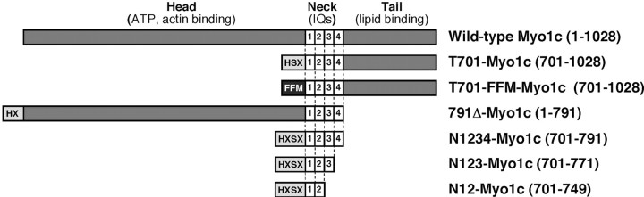
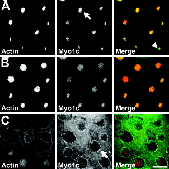
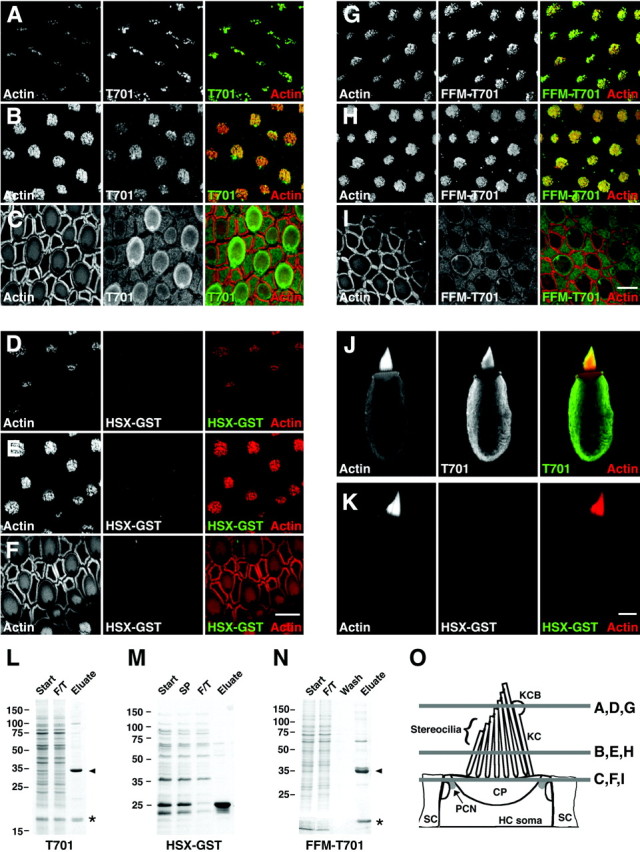
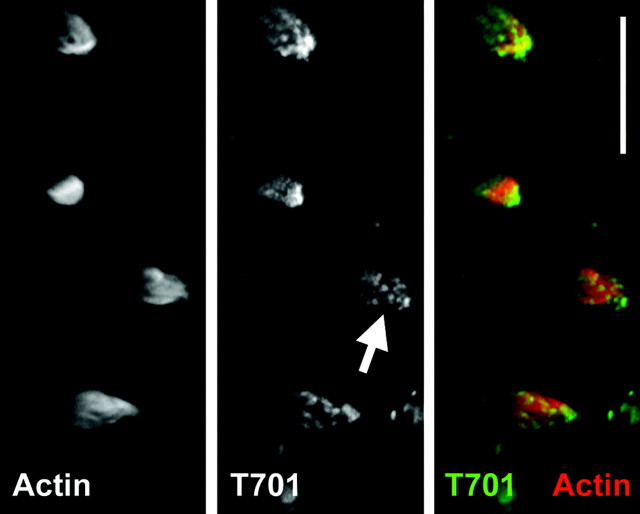
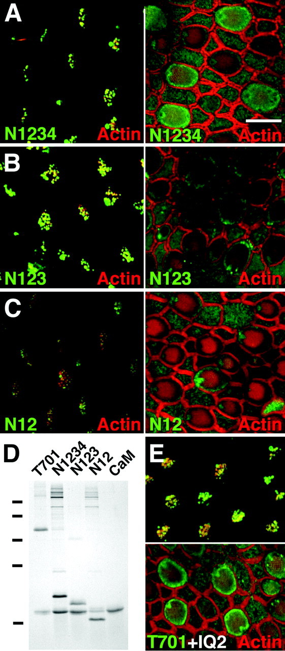
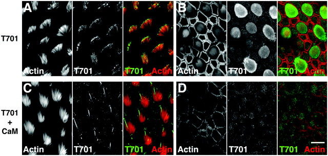
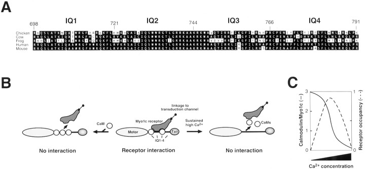
Myosin-1c plays an essential role in adaptation of hair-cell mechanoelectrical transduction. To mediate adaptation, myosin-1c must interact directly or indirectly with other components of the transduction apparatus, including the mechanically gated transduction channel. As a first step toward identifying myosin-1c receptors, we used recombinant myosin-1c fragments to identify specific binding sites in hair cells and to biochemically characterize their interaction with myosin-1c. Myosin-1c fragments bound to tips of hair-cell stereocilia, the location of transduction and adaptation. Surprisingly, this interaction did not depend on the C-terminal tail of myosin-1c, proposed previously to be the receptor-binding site of the molecule. Instead, the interaction of myosin-1c with stereociliary receptors depended on its calmodulin-binding IQ domains. This interaction was blocked by calmodulin, which probably bound to a previously unoccupied IQ domain of myosin-1c. The calcium-sensitive binding of calmodulin to myosin-1c may therefore modulate the interaction of the adaptation motor with other components of the transduction apparatus.


Figures

Fig. 1.
Fig. 1.
Recombinant Myo1c constructs. Myo-1c fragments used in these studies. Shaded regions represent Myo1c sequences, and N-terminal boxes depict epitope tags used for purification and detection. Amino acid residues are indicated inparentheses.
Fig. 2.
Fig. 2.
Localization of Myo1c in the bullfrog sacculus by immunofluorescence. Fluorescence images of a sacculus at three optical-section planes (see Fig. 3O for schematic).Left column, Actin (phalloidin); middle column, Myo1c (R-4280 antibody); right column, combined actin (red) and endogenous Myo1c (green). A, At the tops of hair bundles, Myo1c immunoreactivity is enriched at stereociliary tips (arrow), the kinocilium, and the kinociliary bulb (arrowhead). B, In sterociliary shafts, endogenous Myo1c labeling is present but reduced compared with tip labeling. C, At the apical surface of the sacculus, endogenous Myo1c labeling is strongly enriched along the hair-cell pericuticular necklace (arrow). Scale bar, 10 μm.
Fig. 3.
Fig. 3.
Binding of Myo1c-calmodulin probes to hair-cell receptors. A–I, Left columns, Actin (phalloidin); middlecolumns, epitope-tagged Myo1c or control probe; right columns, combined actin (red) and receptors (green). A–C, T701 binding at stereociliary tips, stereociliary shafts, and pericuticular necklace, respectively. D–F, HSX-GST (negative control) binding at stereociliary tips, stereociliary shafts, and pericuticular necklace, respectively. G–I, Binding of FFM-T701 to hair-cell receptors at stereociliary tips, stereociliary shafts, and pericuticular necklace, respectively. Pericuticular necklace binding is reduced in this particular sample. J, Labeling of isolated hair cell with T701. This cell did not have an intact kinocilium. K, Labeling of an isolated hair cell with HSX-GST. L–N, Coomassie blue-stained SDS-PAGE gels of purified T701, HSX-GST, and FFM-T701. Start, Starting material; SP, soluble proteins;F/T, column flow through; Wash, column wash; Eluate, column eluate. Arrowhead, Myo1c fragments; asterisk, copurifying calmodulin.O, Locations of saccular confocal cross sections shown in A–I. KCB, Kinociliary bulb;KC, kinocilium; HC, hair cell;SC, supporting cell; CP, cuticular plate;PCN, pericuticular necklace. Scale bars: 10 μm (F applies to A–F; Iapplies to G–I; K applies toJ, K).
Fig. 4.
Fig. 4.
Binding of Myo1c–calmodulin probes to isolated, unfixed hair bundles. Hair bundles were isolated in agarose, permeabilized with saponin, and then labeled with phalloidin and T701. No fixation was used. Left, Actin (phalloidin);middle, T701-Myo1c; right, combined actin (red) and T701 (green). Hair-bundle labeling by T701 was pronounced, particularly at stereociliary tips (arrow). Scale bar, 10 μm.
Fig. 5.
Fig. 5.
Binding of Myo1c neck fragments to hair-cell receptors. A–C, Confocal sections of sacculi at hair-bundle tips (left) and pericuticular necklace (right). Green, N1234, N123, or N12 binding; red, actin. N12 labeling of stereociliary tips was diminished compared with N1234 or N123. D, Coomassie-stained SDS-PAGE gel showing purified complexes of calmodulin (CaM) with T701, N1234, N123, and N12. Molecular mass markers of 75, 50, 35, 25, and 15 kDa are indicated to theleft. E, IQ2 peptide (25 μm) does not affect T701 (230 nm) labeling.Top, Sterociliary tips; bottom, pericuticular necklace. Scale bar: A–C, E, 10 μm.
Fig. 6.
Fig. 6.
Calmodulin blocks Myo1c–receptor interaction.Left columns, Actin (phalloidin); middle columns, Myo1c receptors (T701); right columns, combined actin (red) and Myo1c receptors (green). A, B, Labeling of actin and Myo1c receptors (with 230 nm T701) at the tips of stereocilia and pericuticular necklace, respectively.C, D, Simultaneous addition of calmodulin (15 μm) blocks binding of T701 (230 nm) to stereociliary tips and pericuticular necklace but not the kinocilium and kinociliary bulb. Scale bar: A–D, 10 μm.
Fig. 7.
Fig. 7.
Interaction of Myo1c with receptors.A, Alignment of amino acid sequences of the Myo1c neck region from indicated species, with IQ domains indicated. Note the sequence identity of residues 729–748, spanning IQ2 and the beginning of IQ3. B, Model for interaction of Myo1c with receptors. If IQ domains 1–3 are occupied by calmodulin, the receptor cannot bind (left). Calmodulin dissociation from IQ2 permits receptor interaction (middle). If calmodulin dissociates from all IQ domains, receptors cannot bind (right). Calmodulin-bound IQ3 and possibly IQ1 may contribute to the binding site. C, Predicted effects of Ca2+ concentration on the occupancy of Myo1c IQs 1–3 with calmodulin and hence binding to Myo1c receptors.

References

    1. Aruffo A, Stamenkovic I, Melnick M, Underhill CB, Seed B. CD44 is the principal cell surface receptor for hyaluronate. Cell. 1990;61:1303–1313. - PubMed
    1. Assad JA, Hacohen N, Corey DP. Voltage dependence of adaptation and active bundle movement in bullfrog saccular hair cells. Proc Natl Acad Sci USA. 1989;86:2918–2922. - PMC - PubMed
    1. Beningo KA, Lillie SH, Brown SS. The yeast kinesin-related protein Smy1p exerts its effects on the class V myosin Myo2p via a physical interaction. Mol Biol Cell. 2000;11:691–702. - PMC - PubMed
    1. Berg JS, Powell BC, Cheney RE. A millennial myosin census. Mol Biol Cell. 2001;12:780–794. - PMC - PubMed
    1. Bradford MM. A rapid and sensitive method for the quantitation of microgram quantities of protein utilizing the principle of protein-dye binding. Anal Biochem. 1976;72:248–254. - PubMed

Publication types

MeSH terms

LinkOut - more resources