. 2002 Apr 1;22(7):2505-12.
doi: 10.1523/JNEUROSCI.22-07-02505.2002.

Disease-specific human glycine receptor alpha1 subunit causes hyperekplexia phenotype and impaired glycine- and GABA(A)-receptor transmission in transgenic mice

Affiliations

Disease-specific human glycine receptor alpha1 subunit causes hyperekplexia phenotype and impaired glycine- and GABA(A)-receptor transmission in transgenic mice

Lore Becker et al. J Neurosci. .

Abstract

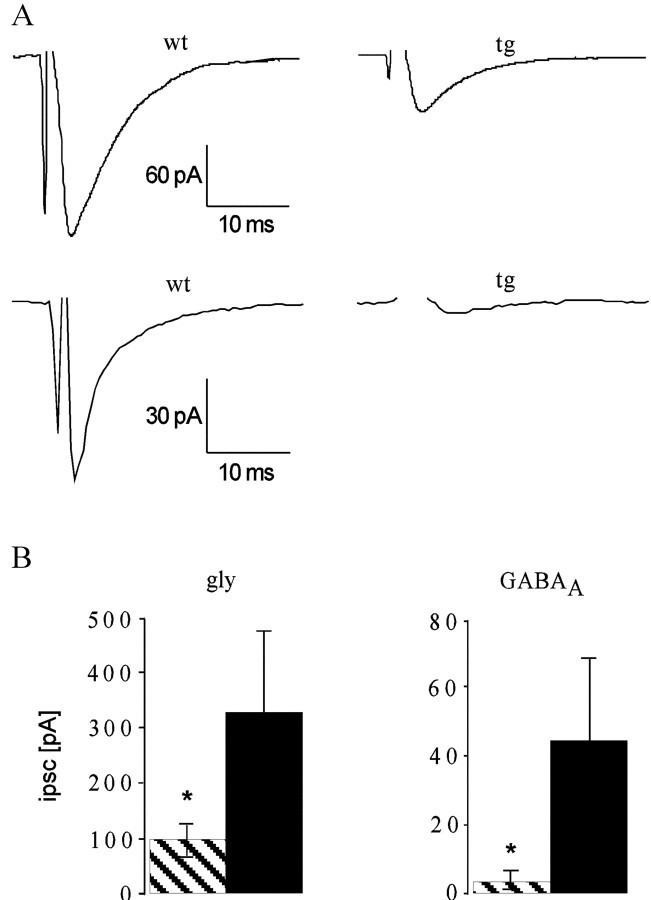
Hereditary hyperekplexia is caused by disinhibition of motoneurons resulting from mutations in the ionotropic receptor for the inhibitory neurotransmitter glycine (GlyR). To study the pathomechanisms involved in vivo, we generated and analyzed transgenic mice expressing the hyperekplexia-specific dominant mutant human GlyR alpha1 subunit 271Q. Tg271Q transgenic mice, in contrast to transgenic animals expressing a wild-type human alpha1 subunit (tg271R), display a dramatic phenotype similar to spontaneous and engineered mouse mutations expressing reduced levels of GlyR. Electrophysiological analysis in the ventral horn of the spinal cord of tg271Q mice revealed a diminished GlyR transmission. Intriguingly, an even larger reduction was found for GABA(A)-receptor-mediated inhibitory transmission, indicating that the expression of this disease gene not only affects the glycinergic system but also leads to a drastic downregulation of the entire postsynaptic inhibition. Therefore, the transgenic mice generated here provide a new animal model of systemic receptor interaction to study inherited and acquired neuromotor deficiencies at different functional levels and to develop novel therapeutic concepts for these diseases.


Figures

Fig. 1.
Fig. 1.
Transgene expression in GlyR transgenic mice is shown. A, Transgene constructs used. The human GlyR cDNAs (open bar) were cloned in a murine genomicThy-1 gene construct in which a region from exon 2 to exon 4 was deleted (Moechars et al., 1996) to confer neuron-specific expression (hatched bars, exon sequences; solid gray bars, intron sequences). The hyperekplexia-specific point mutation is marked by an arrow and anasterisk. B, Semiquantitative RT-PCR of brainstem and spinal cord (sc, black bars) and forebrain (fb,hatched bars) of different mouse lines. The expression of tg271Q-300 in spinal cord was set as 100%. The differences in spinal cord between wt and tg271Q-300 and tg271R-783 mice are significant (asterisks; p < 0.05)C, D, In situ hybridization of sagittal brain sections with GlyR α-specific RNA probes of a wt (C) and a tg271Q-300 (D) animal. E, 3H-strychnine binding to sagittal sections of wt (left), tg271Q-300 (center), and tg271R-783 (right) mice.
Fig. 2.
Fig. 2.
GlyR ligand binding to membranes from the brainstem and spinal cord is shown. A, Specific binding of 3H-strychnine in different genotypes (solid line, wt; dotted line, tg271Q-300; dashed line, tg271R-783). The asterisk at 50 nm marks a significant difference (p < 0.05) between the wt and the transgenic strains. B, C, An 18 nmconcentration of 3H-strychnine was displaced by increasing concentrations of either unlabeled strychnine (B) or glycine (C). Asterisks indicate significance (p < 0.05) D, Displacement of 3H-strychnine with unlabeled ligands on spinal cord sections from different genotypes is indicated; [Note the difference in binding in the tissue of the mutant transgenic animals (tg271Q-300 and tg271Q-382) compared with the wt transgenic (tg271R-783) and the wt line in the presence of 100 μmglycine (bottom).]
Fig. 3.
Fig. 3.
Phenotypic characteristics of tg271Q-300 mice.A, Tremor and disturbed righting when turned to the back. B, Handling-induced tremor, visible at the limbs.C, Hind feet clenching when picked up by the tail.
Fig. 4.
Fig. 4.
Phenotypic analysis of tg271Q-300 animals.A, The time required to right after being turned to the back (n = 65). B, The onset of inducible tremor (n = 42).
Fig. 5.
Fig. 5.
Tremor recordings from transgenic and control mice. Traces of wt (A), and tg271R-783 wt transgenic (B) mice do not show any spastic motor disorder. The tremor in tg271Q-300 mice (D) shows a larger amplitude but a similar tremor frequency compared with homozygous spa mice (E).C, Trace taken from a tg271Q-300 animal during a nontremor period.
Fig. 6.
Fig. 6.
Electrically evoked IPSCs from spinal cord neurons are shown. A, Representative traces from individual measurements on spinal cord neurons of wt (left) and tg271Q-300 (right) mice.Top, Glycinergic component of the IPSC.Bottom, GABAA receptor-mediated IPSC.B, Summary of the electrophysiological data. Mean amplitudes of the glycine-receptor-mediated (left) and GABAA-receptor-mediated IPSCs for tg271Q-300 animals (striped columns) and wt controls (black columns) are shown. In tg271Q-300 animals, the glycinergic component was reduced from −331.2 ± 124.8 pA to −100.1 ± 23.8 pA. The GABAA-receptor-mediated component was −4.1 ± 3.0 pA for tg271Q-300 mice, compared with −45.0 ± 25.4 pA in the controls. Asterisks indicate significance (p < 0.05).
Fig. 7.
Fig. 7.
Autoradiographic studies of brains with benzodiazepine ligands. Distribution of 3H-RO15-4513 binding sites in the presence or absence of flumazenil in brain sections of wt, tg271Q-783, and tg271R-783 transgenic animals are shown.

References

    1. Andrew M, Owen MJ. Hyperekplexia: abnormal startle response due to glycine receptor mutations. Br J Psychiatry. 1997;170:106–108. - PubMed
    1. Becker CM, Hermans-Borgmeyer I, Schmitt B, Betz H. The glycine receptor deficiency of the mutant mouse spastic: evidence for normal glycine receptor structure and localization. J Neurosci. 1986;6:1358–1364. - PMC - PubMed
    1. Becker CM, Schmieden V, Tarroni P, Strasser U, Betz H. Isoform-selective deficit of glycine receptors in the mouse mutant spastic. Neuron. 1992;8:283–289. - PubMed
    1. Becker L, Hartenstein B, Schenkel J, Kuhse J, Betz H, Weiher H. Transient neuromotor phenotype in transgenic spastic mice expressing low levels of glycine receptor β-subunit: an animal model of startle disease. Eur J Neurosci. 2000;12:27–32. - PMC - PubMed
    1. Bekkers JM, Stevens CF. NMDA and non-NMDA receptors are co-localized at individual excitatory synapses in cultured rat hippocampus. Nature. 1989;341:230–233. - PubMed

Publication types

MeSH terms