. 2002 Apr 1;22(7):2590-7.
doi: 10.1523/JNEUROSCI.22-07-02590.2002.

Cdk5/p35 regulates neurotransmitter release through phosphorylation and downregulation of P/Q-type voltage-dependent calcium channel activity

Affiliations

Cdk5/p35 regulates neurotransmitter release through phosphorylation and downregulation of P/Q-type voltage-dependent calcium channel activity

Kazuhito Tomizawa et al. J Neurosci. .

Abstract

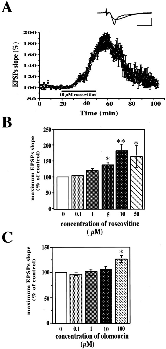
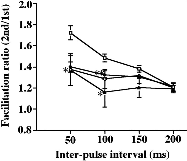
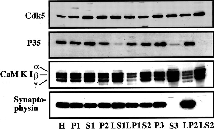
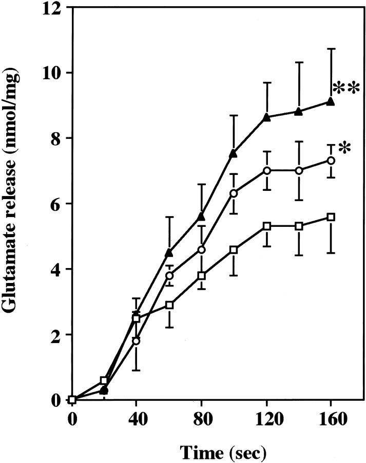
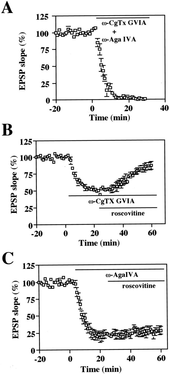
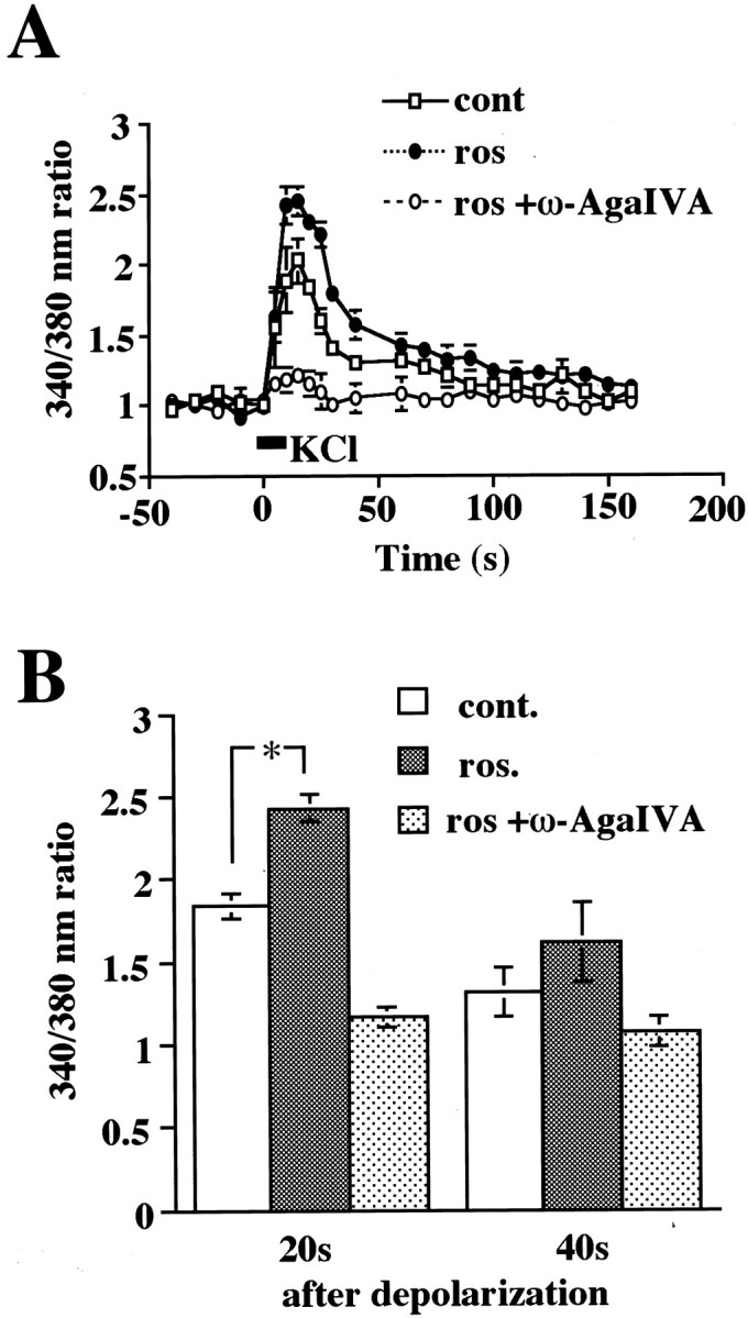
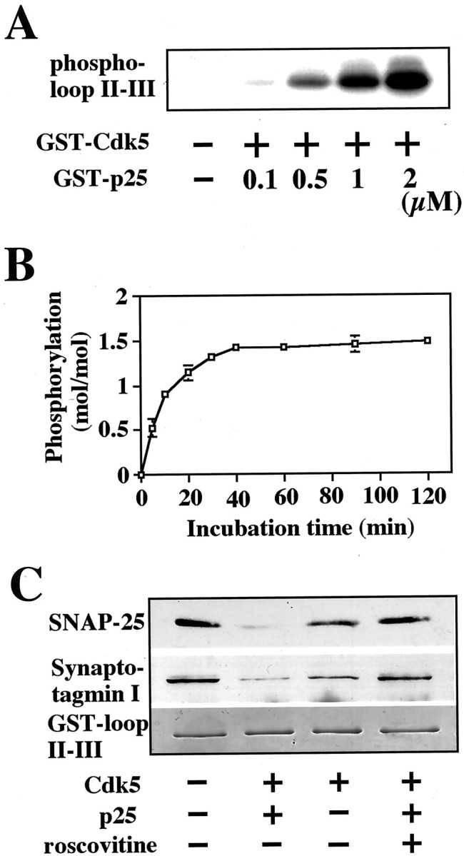
Cyclin-dependent kinase 5 (Cdk5) is a proline-directed serine/threonine kinase with close structural homology to the mitotic Cdks. The complex of Cdk5 and p35, the neuron-specific regulatory subunit of Cdk5, plays important roles in brain development, such as neuronal migration and neurite outgrowth. Moreover, Cdk5 is thought to be involved in the promotion of neurodegeneration in Alzheimer's disease. Cdk5 is abundant in mature neurons; however, its physiological functions in the adult brain are unknown. Here we show that Cdk5/p35 regulates neurotransmitter release in the presynaptic terminal. Both Cdk5 and p35 were abundant in the synaptosomes. Roscovitine, a specific inhibitor of Cdk5 in neurons, induced neurotransmitter release from the synaptosomes in response to membrane depolarization and enhanced the EPSP slopes in rat hippocampal slices. The electrophysiological study using each specific inhibitor of the voltage-dependent calcium channels (VDCCs) and calcium imaging revealed that roscovitine enhanced Ca2+ influx from the P/Q-type VDCC. Moreover, Cdk5/p25 phosphorylated the intracellular loop connecting domains II and III (L(II-III)) between amino acid residues 724 and 981 of isoforms cloned from rat brain of the alpha1A subunit of P/Q-type Ca2+ channels. The phosphorylation inhibited the interaction of L(II-III) with SNAP-25 and synaptotagmin I, which were plasma membrane soluble N-ethylmaleimide-sensitive factor attachment protein (SNAP) receptor (SNARE) proteins and were required for efficient neurotransmitter release. These results strongly suggest that Cdk5/p35 inhibits neurotransmitter release through the phosphorylation of P/Q-type VDCC and downregulation of the channel activity.


Figures

Fig. 1.
Fig. 1.
Cdk5 inhibitors enhanced the field EPSP slope.A, Hippocampal slices were perfused with 10 μm roscovitine for 35 min, and then the slices were washed out by perfusion with ACSF. Inset, Representative field EPSPs before and 40 min after the perfusion of roscovitine are shown. Calibration: 20 msec, 2 mV. B, Dose-dependent effect of roscovitine on maximum EPSPs. To record the maximum EPSP, each concentration of roscovitine was applied until the maximum and stable EPSP was recorded. Each column is the mean ± SEM values of five independent slices. The significance of differences was calculated by the Scheffe's test after ANOVA. *p < 0.05 and **p < 0.01 compared with the control slices (0 μm). C, Dose-dependent effect of olomoucine on the maximum EPSPs. The conditions of drug application were the same as those of roscovitine. Values were significantly different from the control slices (0 μm); *p < 0.05.
Fig. 2.
Fig. 2.
Roscovitine reduced PPF in hippocampal slices. The hippocampal slices were incubated with each concentration of roscovitine (■, 0 μm; ♦, 1 μm; ○, 5 μm; ▴, 10 μm) for 50 min, after which PPF was induced by two pulses of stimulation separated by intervals of 50, 100, 150, and 200 msec. Data show the ratios of the second EPSP slopes and the first EPSP slopes. The significance of difference was calculated by the Scheffe's test after ANOVA. *p< 0.05 and **p < 0.01 compared with the control slices (0 μm).
Fig. 3.
Fig. 3.
Both Cdk5 and p35 were enriched in purified synaptosomes. Western blot analyses for Cdk5, p35, calmodulin kinase I (CaM KI, soluble presynapse fraction marker), and synaptophysin (insoluble presynapse fraction marker) of the rat brain subcellular fractions are shown. Each fraction is shown in Materials and Methods. H, Total homogenate;P2, crude synaptosomes; LP2, membrane fraction of the purified synaptosomes; LS2, cytosolic fraction of the purified synaptosomes.
Fig. 4.
Fig. 4.
The effect of roscovitine on Ca2+-dependent glutamate release from synaptosomes prepared from rat brains in response to membrane depolarization with 1 mm 4-AP (n = 5). Data represent the mean ± SEM values of five independent experiments. The synaptosomes were incubated with each concentration of roscovitine (■, 0 μm; ○, 5 μm; ▴, 10 μm) for 30 min, and then 4-AP was added at 0 sec. Values significantly different from the control synaptosomes (0 μm roscovitine) are indicated (*p < 0.05; **p < 0.01; Scheffe's test).
Fig. 5.
Fig. 5.
The effect of roscovitine on the EPSP slope in the presence of ω-CgTx GVIA and ω-Aga IVA. Hippocampal slices were incubated with 1 μm ω-CgTx GVIA and 0.5 μm ω-Aga IVA (A), 1 μm ω-CgTx GVIA (B), or 0.5 μm ω-Aga IVA (C). Then, 10 μm roscovitine was applied after recording stable EPSP slopes. The drugs were perfused continuously until the end of the experiment.
Fig. 6.
Fig. 6.
Roscovitine enhanced Ca2+influx from P/Q-type VDCCs. A, Primary cultured hippocampal neurons were incubated with ω-CgTx GVIA, Nifedipine, and AP-5 to measure the Ca2+ influx specific from P/Q-type VDCCs for 30 min. Roscovitine was also applied 30 min before depolarization. The cells were exposed with 50 mm KCl for 10 sec, and fura-2 AM fluorescence was recorded using a video image analysis system (AquaCosmos, Hamamatsu Photonics), with excitation alternatively at 340 and 380 nm, and emission at 510 nm (■, control; ●, 5 μm roscovitine; ○, roscovitine and ω-Aga IVA).B, Comparison of Ca2+ influx from P/Q-type VDCCs at 20 and 40 sec after depolarization. Data from 20–30 individual cells were collected per experiment, and ensemble averages were calculated for multiple experiments (n > 30). Data were analyzed using ANOVA followed by planned comparisons of the multiple conditions. *p < 0.01.
Fig. 7.
Fig. 7.
Cdk5/p35-dependent phosphorylation of LII-III of rbA isoform of the α1A subunit of P/Q-type Ca2+ channels and the regulation of the interaction with SNARE proteins. A, Purified GST–LII-III peptides were incubated with GST-Cdk5 or each concentration of GST–p25 at 32°C for 30 min. GST–LII-III peptides were phosphorylated by GST–Cdk5 correlating with GST–p25 concentration. B, Stoichiometry of phosphorylation for GST–LII-III. Polypeptides of GST–LII-III were phosphorylated with GST–Cdk5/p25 for the indicated time periods. C, Phosphorylation of GST–LII-III inhibited the interaction with SNAP-25 and synaptotagmin. Polypeptides of GST–LII-III were incubated with purified Cdk5/p25 or 5 μm roscovitine. Then the complex was incubated with glutathione–Agarose. The glutathione–Agarose was replaced in a 10 ml column. After the column was washed, the solubilized synaptosome protein (300 μg) in HEPES buffer with 20 μmCaCl2 was loaded on the column, and the column was washed. After the protein complexes were collected with glutathione–Agarose, the complex was denatured with boiled SDS-PAGE buffer. The supernatants were then used for Western blot analysis using anti-SNAP-25 antibody, anti-synaptotagmin I antibody, and anti-syntaxin I antibody, respectively. Top panel, Result of Western blot analysis with anti-SNAP-25 antibody. Middle panel, Result of Western blot analysis with anti-synaptotagmin I antibody. Bottom panel, After the complex was collected with glutathione–Agarose, the proteins were separated in SDS-PAGE gel. Then, the gel was stained by Coomassie blue, and the polypeptides of GST–LII-III were visualized.

References

    1. Bibb JA, Snyder GL, Nishi A, Yan Z, Meijer L, Fienberg AA, Tsai LH, Kwon YT, Girault JA, Czernik AJ, Huganir RL, Hemmings HC, Jr, Nairn AC, Greengard P. Phosphorylation of DARPP-32 by Cdk5 modulates dopamine signalling in neurons. Nature. 1999;402:669–671. - PubMed
    1. Catterall WA. Structure and function of neuronal Ca2+ channels and their role in neurotransmitter release. Cell Calcium. 1998;24:307–323. - PubMed
    1. Chae T, Kwon YT, Bronson R, Dikkes P, Li E, Tsai LH. Mice lacking p35, a neuronal specific activator of Cdk5, display cortical lamination defects, seizures, and adult lethality. Neuron. 1997;18:29–42. - PubMed
    1. Dunlap K, Luebke JI, Turner TJ. Exocytotic Ca2+ channels in mammalian central neurons. Trends Neurosci. 1995;18:89–98. - PubMed
    1. Floyd SR, Porro EB, Slepnev VI, Ochoa GC, Tsai LH, De Camilli P. Amphiphysin binds the cdk5 regulatory subunit p35 and is phosphorylated by cdk5 and cdc2. J Biol Chem. 2001;276:8104–8110. - PubMed

Publication types

MeSH terms