

Imaging attentional modulation of pain in the periaqueductal gray in humans
- PMID: 11923440
- PMCID: PMC6758341
- DOI: 10.1523/JNEUROSCI.22-07-02748.2002
Imaging attentional modulation of pain in the periaqueductal gray in humans
Abstract
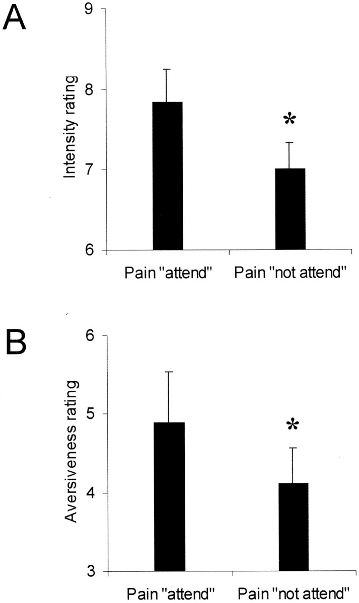
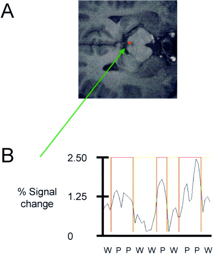
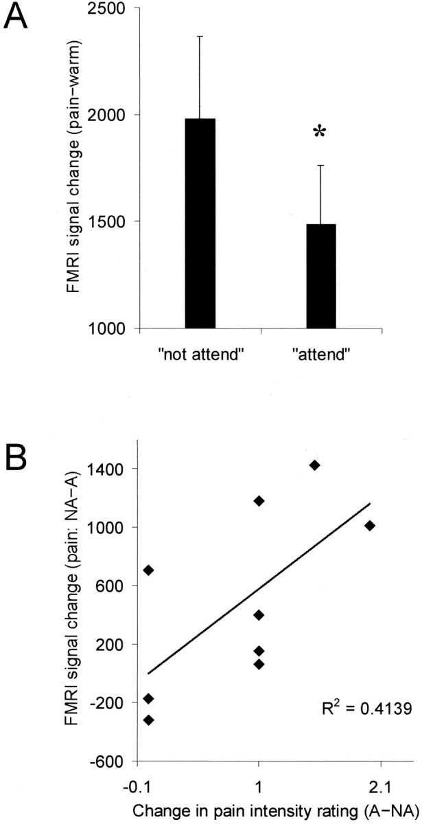
Pain is an unpleasant sensory and emotional experience usually triggered by stimulation of peripheral nerves and often associated with actual or potential tissue damage. It is well known that pain perception for patients and normal subjects can be modulated by psychological factors, such as attention, stress, and arousal. Our understanding of how this modulation occurs at a neuroanatomical level is poor. Here we neuroanatomically defined a key area in the network of brain regions active in response to pain that is modulated by attention to the painful stimulus. High-resolution functional magnetic resonance imaging was used to define brain activation to painful heat stimulation applied to the hand of nine normal subjects within the periaqueductal gray region. Subjects were asked to either focus on or distract themselves from the painful stimuli, which were cued using colored lights. During the distraction condition, subjects rated the pain intensity as significantly lower compared with when they attended to the stimulus. Activation in the periaqueductal gray was significantly increased during the distraction condition, and the total increase in activation was predictive of changes in perceived intensity. This provides direct evidence supporting the notion that the periaqueductal gray is a site for higher cortical control of pain modulation in humans.
Figures
References
-
- Akil H, Mayer DJ, Liebeskind JC. Antagonism of stimulation-produced analgesia by naloxone, a narcotic antagonist. Science. 1976;191:961–962. - PubMed
-
- Albe-Fessard D, Berkley KJ, Kruger L, Ralston HJ, III, Willis WD., Jr Diencephalic mechanisms of pain sensation. Brain Res. 1985;3:217–296. - PubMed
-
- Basbaum AI, Fields HL. Endogenous pain control systems: brainstem spinal pathways and endorphin circuitry. Annu Rev Neurosci. 1984;7:309–338. - PubMed
-
- Baskin DS, Mehler WR, Hosobuchi Y, Richardson DE, Adams JE, Flitter MA. Autopsy analysis of the safety, efficacy and cartography of electrical stimulation of the central gray in humans. Brain Res. 1986;371:231–236. - PubMed
Publication types
MeSH terms
LinkOut - more resources
Full Text Sources
Other Literature Sources
Medical