. 2002 Apr;30(4):401-5.
doi: 10.1038/ng838. Epub 2002 Feb 4.

Microtubule-associated protein 1A is a modifier of tubby hearing (moth1)

Affiliations

Microtubule-associated protein 1A is a modifier of tubby hearing (moth1)

Akihiro Ikeda et al. Nat Genet. 2002 Apr.

Abstract

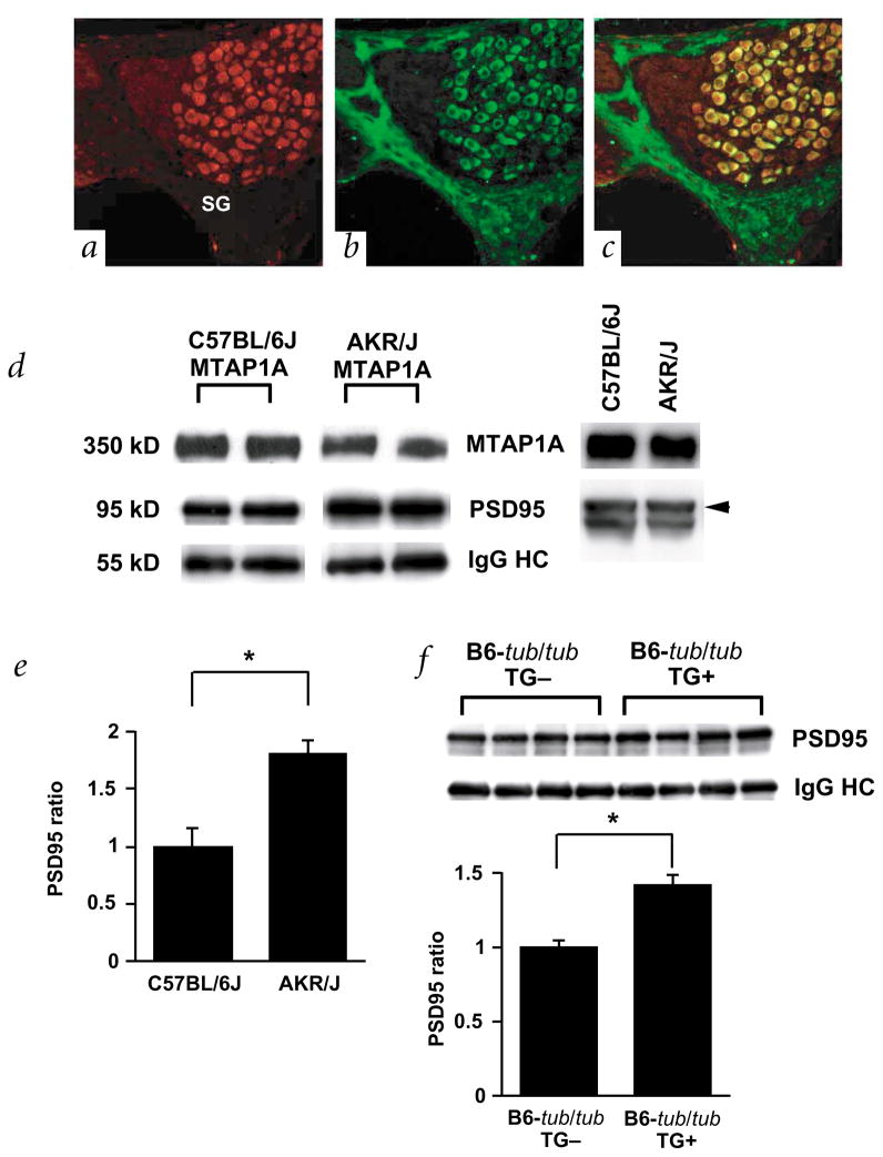
Once a mutation in the gene tub was identified as the cause of obesity, retinal degeneration and hearing loss in tubby mice, it became increasingly evident that the members of the tub gene family (tulps) influence maintenance and function of the neuronal cell lineage. Suggested molecular functions of tubby-like proteins include roles in vesicular trafficking, mediation of insulin signaling and gene transcription. The mechanisms through which tub functions in neurons, however, have yet to be elucidated. Here we report the positional cloning of an auditory quantitative trait locus (QTL), the modifier of tubby hearing 1 gene (moth1), whose wildtype alleles from strains AKR/J, CAST/Ei and 129P2/OlaHsd protect tubby mice from hearing loss. Through a transgenic rescue experiment, we verified that sequence polymorphisms in the neuron-specific microtubule-associated protein 1a gene (Mtap1a) observed in the susceptible strain C57BL/6J (B6) are crucial for the hearing-loss phenotype. We also show that these polymorphisms change the binding efficiency of MTAP1A to postsynaptic density molecule 95 (PSD95), a core component in the cytoarchitecture of synapses. This indicates that at least some of the observed polymorphisms are functionally important and that the hearing loss in C57BL/6J-tub/tub (B6-tub/tub) mice may be caused by impaired protein interactions involving MTAP1A. We therefore propose that tub may be associated with synaptic function in neuronal cells.


Figures

Fig. 1
Fig. 1
Generation of physical contig and gene expression map. a, Fine-structure map of the mothl locus compiled by examining 1,780 meioses from B6-tub/tub intercrosses with AKR/J and CAST/Ei. The genetic size of the critical region is 0.17 ± 0.10 cM. The number of chromosomes sharing the corresponding haplo-types are indicated above each column. b, Representative click ABR thresholds, of offspring from two recombinant individuals, that were key in determining the minimal genetic region of the moth1 locus. Genotypes of recombinant progeny are indicated (top) and corresponding ABR thresholds are shown below (mean ± s.e.m.). The number of progeny tested in each recombinant line is in parentheses. c, Schematic representation of the genetic and physical maps of four overlapping P1/BAC large-insert clones. Transcripts derived by direct cDNA selection and from end-sequencing of clones as well as STS markers are localized on the map. The approximate location of EST AA625130 is marked by a dotted line.
Fig. 2
Fig. 2
Domain structure of MTAP1A and positions of polymorphisms. The location of the light chain 2 (LC2), membrane-associated guanylate kinases (MAGUKs) binding domain and the Ala-Pro (AP) repeat are indicated. Filled circles indicate conservative replacement, and asterisks nonconservative replacement, of amino acids. Note that an AP repeat-length polymorphism is seen between susceptible and protective alleles.
Fig. 3
Fig. 3
Transgenic rescue of hearing impairment in B6-tub/tub by the 129P2/OlaHsd allele of Mtap1a. a, Transgenic rescue construct. Primers for amplification of the GCTCAA repeat used to distinguish between the B6 allele and the transgene derived from 129P2/OlaHsd alleles are indicated. b, Detection of transgene-positive (Tg+) mice by PCR. The size of the resultant PCR products are 274 bp for 129P2/OlaHsd and 214 bp for B6. c, Expression of the transgene-derived Mtap1a in the cochlea as detected by RT–PCR with the primers used for genotyping (the third band which appears under the transgene-derived 274 bp band represents the heteroduplex between the B6 and 129P2/OlaHsd alleles). d, ABR thresholds in C57BL/6-tub/tub-Tg+ mice. The mean ± s.e.m. of the sound pressure threshold is shown for each stimulus and genotype. Five or six mice at 5–6 wk were tested for each genotype. Threshold levels with different superscripts are significantly different (P<0.01).
Fig. 4
Fig. 4
Localization and characterization of MTAP1A. ac, Immunofluorescence micrographs of the spiral ganglion cells in the organ of corti after double-labeling with antibodies against TUB (red, a) and MTAP1A (green, b). Panel c shows a merged version of a and b. Note that TUB is expressed in both the cytosol and nucleus and that TUB and MTAP1A are co-localized in the cytosol of the spiral ganglion cells (yellow). d, Association of MTAP1A derived from B6 (left) and AKR/J (right) with PSD95 in the cerebellum. Immunoprecipitation with anti-MTAP1A followed by immunoblotting with anti-MTAP1A and anti-PSD95 reveals that the amount of PSD95 precipitated by equal amounts of anti-MTAP1A antibody is higher in AKR/J than B6 (left panel). Immunoblot analysis (right panel) shows that the level of protein expression of both MTAP1A and PSD95 is the same in the cerebellum of B6 and AKR/J mice. Levels of immunoprecipitates were determined for each strain using the same membrane. e, Measurement of the relative amount of immunoprecipitated PSD95 in four individual animals each of strains B6 (n=4) and AKR/J (n=4). Levels were normalized to the level of IgG heavy chain, and the B6 levels were arbitrarily set to 1. The mean ± s.e.m. is represented by a bar (asterisk indicates P<0.05). f, Association of MTAP1A with PSD95 in B6-tub/tub and B6-tub/tub-Tg+ mice. An immunoblot for PSD95 co-precipitated with MTAP1A in each animal (n=4 for each strain) is shown (top panel). Measurement of the relative amount of immunoprecipitated PSD95 in four individual animals of each strain (bottom panel). Levels were normalized to the level of IgG heavy chain, and the B6-tub/tub mice levels were arbitrarily set to 1. The mean ± s.e.m. is represented by a bar (asterisk indicates P<0.05).

Comment in

  • Putting tubby on the MAP.
    Grant SG. Grant SG. Nat Genet. 2002 Apr;30(4):347-8. doi: 10.1038/ng0402-347. Nat Genet. 2002. PMID: 11925554 No abstract available.

References

    1. Noben-Trauth K, Naggert JK, North MA, Nishina PM. A candidate gene for the mouse mutation tubby. Nature. 1996;380:534–538. - PubMed
    1. Kleyn PW, et al. Identification and characterization of the mouse obesity gene tubby: a member of a novel gene family. Cell. 1996;85:281–290. - PubMed
    1. Ikeda S, et al. Cell specific expression of tubby gene family members in the retina. Invest Ophthalmol Vis Sci. 1999;40:2706–2712. - PubMed
    1. Hagstrom SA, Duyao M, North MA, Li T. Retinal degeneration in tulp1−/− mice: vesicular accumulation in the interphotoreceptor matrix. Invest Ophthalmol Vis Sci. 1999;40:2795–2802. - PubMed
    1. Ikeda S, et al. Retinal degeneration but not obesity is observed in null mutants of the tubby-like protein 1 gene. Hum Mol Genet. 2000;22:155–163. - PubMed

Publication types

MeSH terms