. 2002 Apr 2;21(7):1524-34.
doi: 10.1093/emboj/21.7.1524.

CLAC: a novel Alzheimer amyloid plaque component derived from a transmembrane precursor, CLAC-P/collagen type XXV

Affiliations

CLAC: a novel Alzheimer amyloid plaque component derived from a transmembrane precursor, CLAC-P/collagen type XXV

Tadafumi Hashimoto et al. EMBO J. .

Abstract

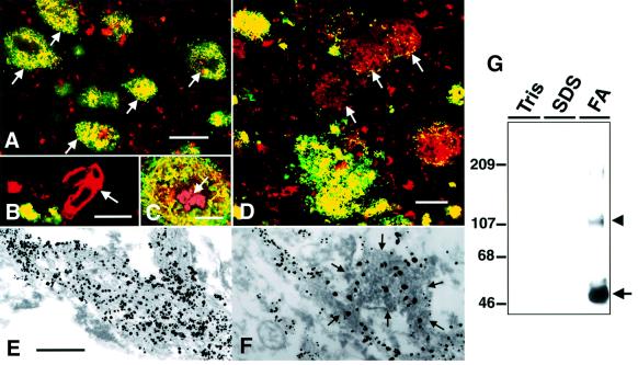
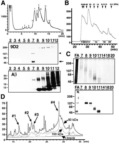
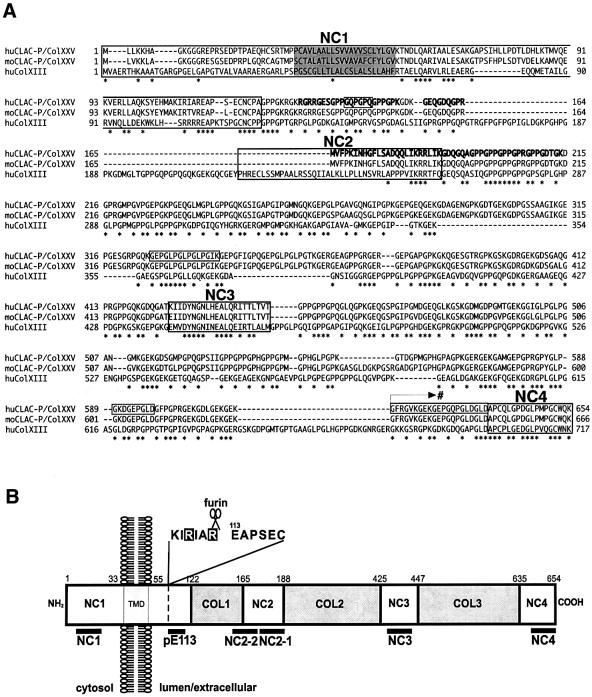
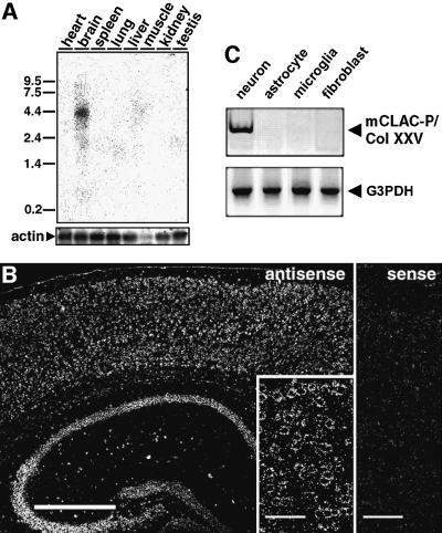
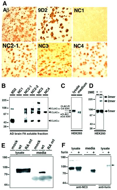
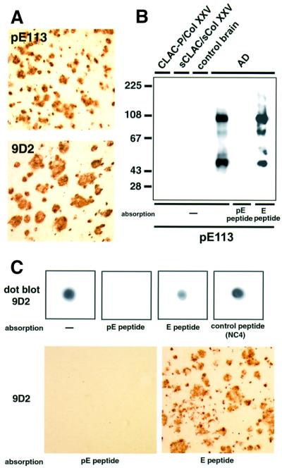
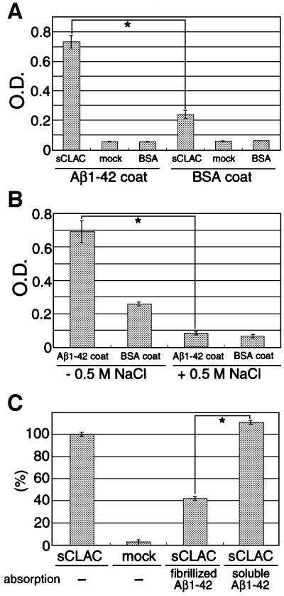
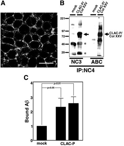
We raised monoclonal antibodies against senile plaque (SP) amyloid and obtained a clone 9D2, which labeled amyloid fibrils in SPs and reacted with approximately 50/100 kDa polypeptides in Alzheimer's disease (AD) brains. We purified the 9D2 antigens and cloned a cDNA encoding its precursor, which was a novel type II transmembrane protein specifically expressed in neurons. This precursor harbored three collagen-like Gly-X-Y repeat motifs and was partially homologous to collagen type XIII. Thus, we named the 9D2 antigen as CLAC (collagen-like Alzheimer amyloid plaque component), and its precursor as CLAC-P/collagen type XXV. The extracellular domain of CLAC-P/collagen type XXV was secreted by furin convertase, and the N-terminus of CLAC deposited in AD brains was pyroglutamate modified. Both secreted and membrane-tethered forms of CLAC-P/collagen type XXV specifically bound to fibrillized Abeta, implicating these proteins in beta-amyloidogenesis and neuronal degeneration in AD.


Figures

None
Fig. 1. Immunohistochemical and biochemical characterization of mAb 9D2. (A) SPs in the frontal cortex of an AD brain doubly immunolabeled by mAb 9D2 (green) and anti-AβN3(pyroGlu) (red). Note that all SPs show areas of homogenous yellow immunofluorescence (arrows), indicating that Aβ and 9D2 antigen are both present. (B) Aβ-positive amyloid angiopathy (arrow; red) in AD cortex is not labeled by mAb 9D2. (C) The core portion of a typical SP (arrow) often lacks 9D2 immunoreactivity (9D2-IR). (D) A subset of Aβ-positive SPs in AD neocortex lack 9D2-IR (arrows, red) although a few dot-like areas of 9D2-IR are present in some SPs. Scale bars in (A–D) are equivalent to 100, 50, 20 and 50 µm, respectively. (E) Immunoelectron microscopic observation of amyloid bundles in SPs of AD neocortex labeled by mAb 9D2. Thick bundles of amyloid fibrils are decorated by coarse particles of 1 nm gold enhanced by silver intensification. The scale bar in (E) is equivalent to 400 nm in (E) and (F). (F) Amyloid fibrils doubly immunolabeled by anti-AβN3(pyroGlu) (1 nm gold particles with silver intensification) and mAb 9D2 (immunoperoxidase labeling). Note that 9D2/immunoperoxidase-positive amyloid bundles (encircled by arrows) were also Aβ-positive (decorated by black particles), indicating that 9D2- and Aβ-IRs are co-localized. (G) Immunoblot analysis of 9D2-positive polypeptides in AD brains. Frozen AD cortices were extracted sequentially by Tris saline (Tris), 2% SDS and 70% formic acid (FA). Note that 9D2-positive polypeptides were extracted exclusively in the FA-soluble fraction as ∼50 (arrow) and ∼100–110 kDa (arrowhead) proteins. Molecular mass standards are shown in kilodaltons.
None
Fig. 2. Purification of 9D2 antigen polypeptide from AD brains. (A) Separation of 9D2 antigen from Aβ in formic acid extracts of AD brains by RP-HPLC. Peaks 2–12 shown in the elution profile (upper panel) were analyzed by immunoblotting with 9D2 (middle panel) or BAN50 (anti-Aβ; lower panel). 9D2-positive ∼50 (arrow) and ∼100–110 kDa (arrowhead) polypeptides were separated in fraction 7, whereas monomeric (∼4 kDa) or oligomeric forms of Aβ were eluted in fractions 8–12. (B) Separation of 9D2 antigen by size exclusion chromatography. The elution profile of fraction 7 in (A) is shown with fraction numbers. Elution positions of molecular mass standard proteins are shown above the panel. (C) Silver-stained gel replica of fractions in (B) separated by SDS–PAGE (upper panel). FA: fraction 7 of formic acid extracts in (A) prior to gel filtration. Immunoblot analysis of fractions 7–20 in (B) with 9D2 (lower panel). Molecular mass standards are shown in kilodaltons. (D) Separation of API-digested peptide fragments derived from ∼50 and ∼100 kDa 9D2 antigen polypeptides by RP-HPLC. Fractions 1–4 that gave partial amino acid sequences of CLAC protein are indicated by arrows. The ordinates in (A) (upper panel), (B) and (D) are shown in milliabsorbance (mA) at 215 nm.
None
Fig. 3. Amino acid sequence, domain structure and topology of CLAC-P/Col XXV. (A) Predicted sequence of human (upper lane) and mouse (middle lane) CLAC-P/Col XXV, as well as of human Col XIII (lower lane). Peptide fragments identified by amino acid sequencing (peaks 1–4 in Figure 2D) are shown in bold. Four non-collagenous domains are boxed with the names of NC1–NC4, and a putative transmembrane domain is shaded. Amino acid residues that undergo alternative splicing in variants of human CLAC-P/Col XXV (i.e. residues 141–146, 326–340, 589–597 and 616–636) are also boxed, and an arrow with # indicates the starting point of a C-terminal splice variant that replaces the C-terminus including the NC4 domain by the amino acid sequence: VTSPSQHVPCLILLLLSALLFSLCDSI (DDBJ/EMBL/GenBank accession No. AF293341; registered as CLAC-P type II). Amino acid residues identical among the three molecular species are marked by asterisks. (B) Schematic representation of the domain structure and topology of human CLAC-P/Col XXV. The NC1 domain composed of cytoplasmic, transmembrane (TMD) and extracellular portions, three extracellular NC domains (NC2–NC4) and the three collagenous domains (COL1–COL3) are shown with amino acid residue numbers. The locations of the epitopes of antibodies used in this study and a putative furin cleavage site within NC1 are shown below and above the column, respectively.
None
Fig. 4. mRNA expression patterns of CLAC-P/Col XXV. (A) Northern blot analysis of CLAC-P/Col XXV mRNA expression in various mouse tissues. The blots were rehybridized using the actin probe as a control of mRNA loading. Standards are shown in kilobases. (BIn situ hybridization of CLAC-P/Col XXV mRNA in mouse cerebral neocortex and hippocampus (left, antisense probe), and a higher magnification of cerebral neocortex showing neuronal localization of signals (inset). Hybridization with a sense probe gave no specific signals (cerebral neocortex, right). Scale bars in (B) are equivalent to 500 µm (left panel) and 50 µm (inset and right panel). (C) RT–PCR analysis of expression of CLAC-P/Col XXV in cell type-specific primary cultures from mouse brains, showing neuron-specific expression of CLAC-P/Col XXV (upper panel). Expression of G3PDH as a control is shown in the lower panel.
None
Fig. 5. Characterization of CLAC polypeptide deposited in AD brains and CLAC-P/Col XXV expressed in HEK293 cells, and secretion of sCLAC/sCol XXV by furin convertase. (A) Immunohistochemistry of AD neocortex stained by anti-AβN3(pyroGlu), 9D2 and anti-CLAC-P/Col XXV antibodies. (B) Immunoblot analysis of formic acid extracts of AD brains probed by mAb 9D2 and anti-CLAC-P/Col XXV antibodies. Note that anti-NC2-1, NC2-2 and NC3 label a set of polypeptides (CLAC50, CLAC70 and CLAC100), whereas anti-NC4 exclusively labeled CLAC70. (C) Immunoblot analysis of HEK293 cells stably expressing human CLAC-P/Col XXV with anti-NC3. Left lane, cell lysate; right lane, culture media. Note that sCLAC/sCol XXV in culture media co-migrates with CLAC70 at ∼70 kDa. Molecular mass standards are common to (B) and (C). (D) Formation of trimers (3mer) and dimers (2mer) of CLAC-P/Col XXV under non-reducing conditions. Cell lysates of HEK293 cells stably transfected with human CLAC-P/Col XXV were analyzed by immunoblotting with anti-NC3 with (left lane) or without (right lane) 2-mercaptoethanol (2ME) pre-treatment. (E) COS-1 cells were transiently transfected with cDNAs encoding wild-type (wt) or RA mutant (mt) human CLAC-P/Col XXV, the latter being substituted with alanine at two arginine residues, at positions –1 and –4 to the putative furin cleavage site, or with an empty vector (mock). Cell lysate (left panel) and culture media (right panel) were analyzed by immunoblotting with anti-NC3. (F) RPE.40 mutant CHO cells that lack furin were stably transfected with human CLAC-P/Col XXV. Lysates and culture media from these stable cells with (+) or without (–) transient co-transfection of mouse furin were analyzed by immunoblotting with anti-NC3 (lysate and media, left panel) or anti-furin (lysate, right panel; the arrow shows transfected furin polypeptide). Molecular mass standards are shown in kilodaltons.
None
Fig. 6. The N-terminus of CLAC deposited in AD brains undergoes pyroglutamate modification. (A) Immunolabeling of SP in the frontal neocortex of AD brains with anti-pE113 (upper panel) and 9D2 (lower panel). (B) Immunoblot analysis of formic acid extracts of AD and control brains, as well as of CLAC-P/Col XXV and sCLAC/sCol XXV in HEK293 cells stably expressing CLAC-P/Col XXV, with anti-pE113. Note that pre-absorption of anti-pE113 with an immunogen peptide (pE peptide), but not with E peptide, abolished positive immunoreaction of CLAC. (C) Characterization of 9D2 as an N-terminal pyroglutamate modification-specific antibody. Dot blot analysis of formic acid extracts (upper panels) or tissue sections (lower panels) of AD brains by 9D2, showing abolition of positive reaction by absorption with pE peptide, but not with E peptide or irrelevant control peptides.
None
Fig. 7. Binding of sCLAC/sCol XXV to Aβ. (A) Bound sCLAC on multi-well plates pre-coated with fibrillized Aβ1–42 or BSA after incubation with conditioned media from HEK293 cells stably transfected with CLAC-P/Col XXV (sCLAC), or that from mock-transfected cells (mock) or BSA as controls, was detected by incubation with anti-NC3 followed by immunoperoxidase reaction. (B) Binding of sCLAC/sCol XXV onto multi-well plates pre-coated with Aβ1–42 or BSA after incubation with conditioned media containing sCLAC/sCol XXV in the presence (right lanes) or absence (left lanes) of 0.5 M NaCl. Mean optical densities ± SD in four independent experiments are shown and *P <0.01 by Student’s t-test in (A) and (B). (C) Binding of sCLAC/sCol XXV to pre-coated Aβ1–42 after incubation with conditioned media of CLAC-P/Col XXV stable (sCLAC) or mock-transfected cells pre-absorbed with fibrillized or soluble Aβ1–42. Percentages ± SD of optical densities in four independent experiments relative to those from incubation of sCLAC/sCol XXV without absorption are shown.
None
Fig. 8. Cell surface expression of CLAC-P/Col XXV and binding of Aβ therewith. (A) Immunofluorescence labeling of HEK293 cells stably transfected with human CLAC-P/Col XXV in a confluent state by anti-NC4. Note that cell surface and subplasmalemmal areas are labeled. Scale bar = 10 µm. (B) Surface biotin labeling of CLAC-P/Col XXV in HEK293 stable cells. After surface labeling of CLAC-P/Col XXV with biotin, cell lysates of CLAC-P/Col XXV stable cells or mock-transfected cells were immunoprecipitated by anti-NC4, followed by immunoblotting with anti-NC3 (left panels), or directly visualized by avidin–biotin complex (ABC). Note that CLAC-P/Col XXV migrating at ∼80 kDa is labeled by both anti-NC3 and ABC (arrow). Asterisks show non-specific reaction of immunoglobulin heavy and light chains. (C) Relative levels of fibrillized Aβ1–42 bound to the cell surface CLAC-P/Col XXV in two HEK293 stable cell lines expressing CLAC-P/Col XXV as assayed by quantitative immunoblotting with BC05 (relative to the level in mock-transfected cells as 1.0). Mean ± SD in six independent experiments for each cell line is shown. *P <0.01 and <0.05, respectively, by Student’s t-test in the two cell lines.

References

    1. Brady M.A.W. and Finlan,M.F. (1990) Radioactive labels: autoradiography and choice of emulsions for in situ hybridization. In Polak,J.M. and McGee,J.O’D. (eds), In situ Hybridization, Principles and Practice. Oxford University Press, Oxford, UK, pp. 31–57.
    1. Bruckner P. and Prockop,D.J. (1981) Proteolytic enzymes as probes for the triple-helical conformation of procollagen. Anal. Biochem., 110, 360–368. - PubMed
    1. Dickson D.W. (1997) The pathogenesis of senile plaques. J. Neuropathol. Exp. Neurol., 56, 321–339. - PubMed
    1. Duff K. et al. (1996) Increased amyloid-β42(43) in brains of mice expressing mutant presenilin 1. Nature, 383, 710–713. - PubMed
    1. Duguay S.J., Lai-Zhang,J. and Steiner,D.F. (1995) Mutational analysis of the insulin-like growth factor I prohormone processing site. J. Biol. Chem., 270, 17566–17574. - PubMed

Publication types

MeSH terms

Associated data