. 2002 Apr 2;21(7):1597-606.
doi: 10.1093/emboj/21.7.1597.

Plasmodium sporozoite invasion into insect and mammalian cells is directed by the same dual binding system

Affiliations

Plasmodium sporozoite invasion into insect and mammalian cells is directed by the same dual binding system

Kai Matuschewski et al. EMBO J. .

Abstract

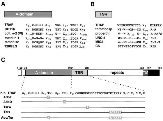
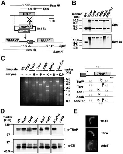
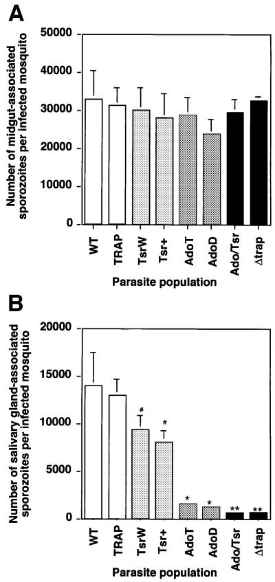
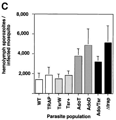
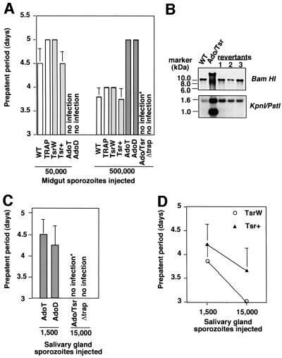
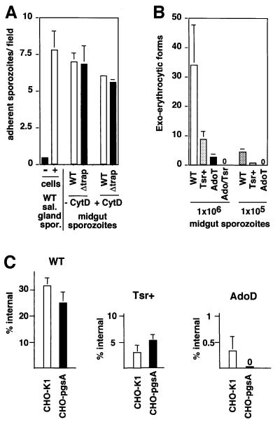
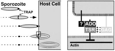
Plasmodium sporozoites, the transmission form of the malaria parasite, successively invade salivary glands in the mosquito vector and the liver in the mammalian host. Sporozoite capacity to invade host cells is mechanistically related to their ability to glide on solid substrates, both activities depending on the transmembrane protein TRAP. Here, we show that loss-of- function mutations in two adhesive modules of the TRAP ectodomain, an integrin-like A-domain and a thrombospondin type I repeat, specifically decrease sporozoite invasion of host cells but do not affect sporozoite gliding and adhesion to cells. Irrespective of the target cell, i.e. in mosquitoes, rodents and cultured human or hamster cells, sporozoites bearing mutations in one module are less invasive, while those bearing mutations in both modules are non-invasive. In Chinese hamster ovary cells, the TRAP modules interact with distinct cell receptors during sporozoite invasion, and thus act as independently active pass keys. As these modules are also present in other members of the TRAP family of proteins in Apicomplexa, they may account for the capacity of these parasites to enter many cell types of phylogenetically distant origins.


Figures

None
Fig. 1. Conserved residues in A-domain- and TSR-containing proteins, and TRAP variants constructed in this study. (A) Comparison of MIDAS motifs in A-domain-containing proteins (non-exhaustive list). The invariant residues of the MIDAS are highlighted in bold. TRAP, P.berghei thrombospondin-related anonymous protein; CD11b, α-subunit of the human integrin Mac-1; coll. α-2 (VI), α2-chain of human collagen VI (consensus of the three vWA-domains); matrilin-1, mouse cartilage matrix protein (consensus of the two vWA-domains); factor C2, human complement factor C2; T20G5.3, hypothetical Caenorhabditis elegans protein T20G5.3. (B) Comparison of the TSRs in TSR-containing proteins (non-exhaustive list). The central region of TSRs is shown, which includes the conserved tryptophans and the cluster of positive residues. The C-terminal conserved cysteines (between two and four) that participate in disulfide bond formation are not shown. TRAP, P.berghei thrombospondin-related anonymous protein; thrombosp., human thrombospondin (consensus of the three TSRs); properdin, human properdin (consensus of the six TSRs); UNC-5, C.elegans netrin-receptor UNC-5 (consensus of the two TSRs); MIC2, Toxoplasma gondii micronemal protein-2 (consensus of the five TSRs; lower cases = conserved in at least three TSRs); CS, P.berghei circumsporozoite protein. (C) Schematic diagram of the primary structure of P.berghei TRAP and of the mutations generated in its A-domain and TSR. In the A-domain, the conserved Thr126 and Asp157 were changed to alanines to generate mutants AdoT and AdoD, respectively. In the TSR, the conserved Trp244 was changed to alanine to generate mutant TsrW. The lysines and arginines towards the C-terminus of the motif were replaced by alanines, leading to mutant Tsr+. The double mutant Ado/Tsr is mutated both in the A-domain (Thr126→Ala) and the TSR (Lys,Arg256–261Ala). Repeats, species-specific amino-acid repeats; TM, transmembrane domain; black box, cyto plasmic domain.
None
Fig. 2. Targeted mutagenesis at the TRAP locus. (A) Insertion strategy to generate the mutant parasite clones. The wild-type (WT) TRAP genomic locus is targeted with an EcoRI (E)-linearized plasmid containing a 3′ truncated TRAP open reading frame and the corresponding base pair substitutions (TRAP*Δ3′) as well as the mutated dhfr/ts gene for selection with pyrimethamine. Upon a single cross-over event the region of homology is duplicated and the mutation is placed in the second, full-length and expressed copy of TRAP (TRAP*). The first copy lacks the 3′ part of TRAP (TRAPΔ3′) as well as downstream regulatory sequences. The restriction fragments generated by SpeI (that cuts once in the plasmid) or BamHI (that does not cut in the plasmid) in WT TRAP and the expected recombinant locus are shown as lines with their predicted sizes. (B) Genomic Southern hybridization. A successful integration event of the targeting plasmid at the TRAP locus is verified by the presence of an additional SpeI fragment of the size of the targeting plasmid (10.5 kbp) as well as the corresponding increase in the size of the BamHI fragment. Note that the clone AdoD contains a tandem integration of two targeting plasmids, increasing the BamHI fragment from 20 to 30.5 kbp. (C) PCR analysis of the TRAP loci of WT and clonal parasite populations. The primers used for specific amplification of the last copy of TRAP, the only one to be full-length and expressed, were within the dhfr/ts resistance marker (forward primer) and the 3′ untranslated region (UTR) of TRAP (reverse primer) and are indicated by arrows. The predicted 3.8 kbp product was amplified from all parasite clones. Restriction digestion of the PCR products confirmed the presence of the corresponding mutations. Abbreviations: H, HindIII; P, PstI; K, KpnI; N, NgoMIV. Numbers indicate the size of the fragments on either side of the restriction site (in kbp). (D) Western blot analysis of sporozoite extracts. Extracts from WT or mutant, midgut sporozoites (∼100 000) were separated on a 8% SDS gel and probed with either a polyclonal serum directed against the P.berghei TRAP repeats, or a monoclonal anti-CS antibody to confirm that similar amounts of sporozoite extracts were loaded in each lane. The nature of the low molecular weight TRAP bands, as well as the reason for their changing ratios (in the WT and mutants), remains unknown. (E) Immunofluores cence stainings of salivary gland sporozoites with anti-TRAP and anti-CS antibodies. Sporozoites in mutant populations show the typical focal staining of TRAP (white) at the sporozoite apical end and the even surface distribution of CS (grey). Note also the deposit of TRAP and CS on the glass slide after gliding (shown for the AdoT parasite).
None
Fig. 3. The TRAP A-domain and TSR are important for sporozoite invasion into A.stephensi salivary glands. Represented in all graphs are the mean numbers of sporozoites (+SEM) calculated from four counts, each from an independent mosquito feeding experiment. In each population, an average of 250 mosquitoes from a minimum of four independent feeding experiments were examined. (A) Midgut sporozoites per infected mosquito at day 14 post-feeding. The variations between the various parasite populations were not statistically significant (P >0.07). (B) Salivary gland sporozoites (attached to, and within the glands) per infected mosquito at day 18 post-feeding. #, the one-tailed P values for Tsr mutants were significantly lower than the mean value for the TRAP control (TsrW: 0.024; Tsr+: 0.028); *, the mean numbers of Ado mutants were significantly lower than the mean value of the TRAP control (P <0.001); **, the mean values of the Ado/Tsr and the Δtrap mutants were significantly lower than the mean value of the Ado mutants (P <0.01). (C) Hemolymph sporozoites per infected mosquito at day 16 post-feeding. Note an increase in the numbers of hemolymph sporozoites in parasite populations that are blocked in salivary gland invasion.
None
Fig. 4. Sporozoite infectivity to the mammalian host. (A) Infectivity to rats of midgut sporozoites. Sporozoites were isolated from midguts of infected mosquitoes and intravenously injected into Sprague/Dawley rats. Shown is the average prepatent period (in days) after injection of 50 000 and 500 000 sporozoites. Ado sporozoites were less infective than TRAP sporozoites, while Ado/Tsr sporozoites were not infective. One rat injected with Ado/Tsr sporozoites became infected (marked with *); however, the resulting erythrocytic stages were revertants [see (B)]. Experiments were carried out in quadruplicate. (B) Genomic Southern hybridization of parasite red blood cell stages obtained after injection of Ado/Tsr sporozoites. Three parasite populations were obtained in rats, after injection of (i) 500 000 midgut sporozoites (one out of four experiments), (ii) 15 000 salivary gland sporozoites (one out of four experiments), and (iii) natural feeding of 100 infected mosquitoes on a rat (one out of four experiments). Parasites were revertants, i.e. arose after excision of the integration plasmid, as shown by the BamHI digestion, and had not retained the mutation in the TRAP gene, as shown by the KpnI–PstI double digestion. (C) Ado/Tsr salivary gland sporozoites are not infective. While injection of 1500 Ado (T or D) sporozoites is sufficient to cause red blood cell infection in rats, injection of 15 000 Ado/Tsr sporozoites does not infect rats. Experiments were carried out in duplicate with three replicas each. *, one rat became infected; however, the resulting erythrocytic stages were revertants [see (B), revertant 2]. (D) Tsr+ salivary gland sporozoites are less infective to rats than TsrW sporozoites after intravenous injection. Infectivity to rodents of Tsr+ and TsrW populations can be compared using salivary gland sporozoites, because these two populations have similar salivary gland infection rates. The prepatent period of infection is shown as a function of the numbers of sporozoites injected. Values are the mean PP from six experiments, each carried out in duplicate. The Tsr+ mutation leads to a consistent delay of patency, corresponding to a 5- to 10-fold decrease in infective capacity.
None
Fig. 5. The TRAP ectodomain is important for sporozoite invasion of, but not adhesion to, cultured cells. (A) TRAP is dispensable for sporozoite adhesion to HepG2 cells. Midgut sporozoites (50 000) were pre-incubated for 15 min in the presence or absence of 2 µM Cytochalasin D subsequently incubated for 90 min with confluent HepG2 cells, and sporozoites that remained bound to cells after washings were stained with anti-CS antibody. Adhesion is shown as the mean number of bound sporozoites in one microscopic field (400× magnification), from two sets of experiments done in quadruplicate each. (B) The TRAP A-domain and TSR are required for invasion of HepG2 cells. Midgut sporozoites (1 000 000) were added to subconfluent HepG2 cells (multiplicity of infection = 1), and EEF that develop inside cells were immunostained 48 h later. The mean numbers of EEFs from two experiments carried out at least in triplicate each are shown. (C) Cell invasion into WT CHO cells (CHO-K1) and GAG-deficient CHO cells (pgsA). Salivary gland sporozoites were incubated for 90 min with confluent CHO cells (empty bars) or pgsA derivatives (black bars) at a multiplicity of infection of 0.01, and the proportion of internalized sporozoites was assessed by differential immunostaining of extra and intracellular sporozoites (10 000 cells examined for each population). Left, GAG chains are of minor importance for WT sporozoite invasion into CHO cells. Center, TSR+ salivary gland sporozoites enter GAG-deficient cells with similar efficiency to WT cells. Right, AdoD sporozoites invade cultured cells in a GAG-dependent fashion.
None
Fig. 6. Hypothetical model for the role of the two TRAP adhesive modules during Plasmodium sporozoite invasion of host cells. Plasmodium sporozoites reach their target cells through gliding locomotion. Left part, TRAP is capped backwards during gliding motility and host cell invasion. The A-domain MIDAS and the TSR of TRAP are not required for this step. Initial host cell attachment is TRAP independent, but induces a burst of TRAP secretion at the sporozoite tip. Right, host cell invasion requires the formation of a junction between the sporozoite anterior end and the host cell. The A-domain, which binds to an as yet unidentified host cell receptor (?) in a MIDAS-dependent fashion, and the TSR, which recruits glycosaminoglycan chains (GAG) via a charged interaction, presumably act during this step of junction formation. An actin-based motor system then generates force, which is transmitted via the TRAP cytoplasmic tail and exerted on the host–parasite junction. This allows the sporozoite to locomote inside the so-called parasitophorous vacuole.

References

    1. Bernfield M., Gotte,M., Park,P.W., Reizes,O., Fitzgerald,M.L., Lincecum,J. and Zako,M. (1999) Functions of cell surface proteoglycans. Annu. Rev. Biochem., 68, 729–777. - PubMed
    1. Carruthers V.B. and Sibley,L.D. (1999) Mobilization of intracellular calcium stimulates microneme discharge in Toxoplasma gondii. Mol. Microbiol., 31, 421–428. - PubMed
    1. Cerami C., Frevert,U., Sinnis,P., Takacs,B., Clavijo,P., Santos,M.J. and Nussenzweig,V. (1992) The basolateral domain of the hepatocyte plasma membrane bears receptors for the circumsporozoite protein of Plasmodium falciparum sporozoites. Cell, 70, 1021–1033. - PubMed
    1. Dessens J.T., Beetsma,A.L., Dimopoulos,G., Wengelnik,K., Crisanti,A., Kafatos,F.C. and Sinden,R.E. (1999) CTRP is essential for mosquito infection by malaria ookinetes. EMBO J., 18, 6221–6227. - PMC - PubMed
    1. Dobrowolski J.M. and Sibley,L.D. (1996) Toxoplasma invasion of mammalian cells is powered by the actin cytoskeleton of the parasite. Cell, 84, 933–939. - PubMed

Publication types

MeSH terms

Substances