. 2002 Apr 2;21(7):1607-15.
doi: 10.1093/emboj/21.7.1607.

IdeS, a novel streptococcal cysteine proteinase with unique specificity for immunoglobulin G

Affiliations

IdeS, a novel streptococcal cysteine proteinase with unique specificity for immunoglobulin G

Ulrich von Pawel-Rammingen et al. EMBO J. .

Abstract

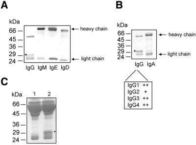
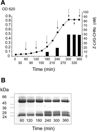
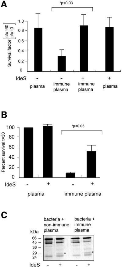
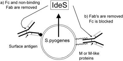
Recent work from several laboratories has demonstrated that proteolytic mechanisms significantly contribute to the molecular interplay between Streptococcus pyogenes, an important human pathogen, and its host. Here we describe the identification, purification and characterization of a novel extracellular cysteine proteinase produced by S.pyogenes. This enzyme, designated IdeS for Immunoglobulin G-degrading enzyme of S.pyogenes, is distinct from the well-characterized streptococcal cysteine proteinase, SpeB, and cleaves human IgG in the hinge region with a high degree of specificity. Thus, other human proteins, including immunoglobulins M, A, D and E, are not degraded by IdeS. The enzyme efficiently cleaves IgG antibodies bound to streptococcal surface structures, thereby inhibiting the killing of S.pyogenes by phagocytic cells. This and additional observations on the distribution and expression of the ideS gene indicate that IdeS represents a novel and significant bacterial virulence determinant, and a potential therapeutic target.


Figures

None
Fig. 1. Cleavage of IgG in S.pyogenes growth medium. (A) Identification of an extra protein band in the culture supernatant of S.pyogenes grown in the presence of human plasma (see asterisk). Lane 1, 5% human plasma in TH. Lane 2, AP1 bacteria grown in TH medium supplemented with human plasma to 10%. Samples were separated on 12% SDS–PAGE and stained with Coomassie Blue. (B) The determined N-terminal amino acid sequence of the 31 kDa protein found in bacterial culture supernatant is boxed. This sequence was identified in the hinge region of human IgG1 and the cleavage site is indicated by an arrow. (C) Human polyclonal IgG was incubated with growth medium (TH) alone (lane 1), with growth medium from strain AP1 lacking (lane 2) or containing E64 (lane 3), with growth medium from the speB-deficient mutant strain AL1 (lane 4), or the mga mutant strain BMJ71 (lane 5). IgG cleavage was analyzed by 12% SDS–PAGE, and the gel was stained with Coomassie Blue. Molecular weight markers are indicated.
None
Fig. 2. Schematic representation of the IdeS protein. The obtained N- terminal sequence (amino acids 30–40), the putative signal sequence (Ss) and an RGD motif (amino acids 214–216) are indicated. The potential catalytic cysteine and histidine residues are designated Cys94 and His224, respectively.
None
Fig. 3. IdeS is a novel streptococcal cysteine proteinase. Purified IdeS or activated SpeB as a control, were separately pre-incubated with proteinase inhibitors prior to incubation with IgG. Cleavage was analyzed by 12% SDS–PAGE. (A) Untreated IdeS. (B–E) IdeS in 1% DMSO (solvent for Z-LVG-CHN2) (B), iodoacetic acid (C), Z-LVG-CHN2 (D) or E64 (E). (F) IgG. (G–I) Untreated SpeB (G), Z-LVG-CHN2 (H) or E64 (I).
None
Fig. 4. IdeS is specific for IgG. (A and B) Human immunoglobulins of different classes were separately incubated with IdeS, followed by SDS–PAGE analysis. Only IgG was degraded by IdeS. All four IgG subclasses are cleaved by IdeS, but IgG2 is more resistant to IdeS cleavage (+). Results from different experiments have been combined. (C) Samples of 5% human plasma in TH (lane 1) or 5% human plasma in TH pre-incubated with IdeS (lane 2) were separated by 12% SDS–PAGE. The asterisk indicates the 31 kDa IgG-derived band.
None
Fig. 5. IgG-cleaving activity generated during streptococcal growth. (A) Strain AP1 was grown in TH medium; supernatant samples were taken at indicated time points (arrows) and IdeS concentrations were determined by adding increasing amounts of Z-LVG-CHN2. Bars indicate the amount inhibitor required to inhibit >90% of enzyme activity. (B) IgG-cleaving activity at different time points as shown by SDS–PAGE.
None
Fig. 6. IdeS cleaves IgG at the bacterial surface and confers resistance against killing of S.pyogenes by phagocytes. (A) AP1 bacteria were incubated with human non-immune or immune plasma, washed, and incubated with or without IdeS. Bacteria were washed and added to RAW264.7 macrophage-like cells. Bacteria were recovered either directly after centrifugation (t0) or after 1 h (t60). The survival rate is shown as number of c.f.u. at 1 h divided by the number of c.f.u. at time zero. Bars indicate the mean values +SEM of at least three different experiments. (B) Bacteria and neutrophils were mixed and incubated at 37°C for various periods of time. Samples were withdrawn and phagocytosis was stopped by putting samples on ice. The bacterial survival rate is shown as number of c.f.u. at 30 min divided by the number of c.f.u. at time zero. The ratio obtained with AP1 treated with non-immune plasma was arbitrarily set to 100%. All other ratios are correlated to this number. Bars indicate the mean values +SEM. (C) AP1 bacteria were incubated with human non-immune or immune plasma, and incubated with IdeS or buffer control. Plasma proteins bound to the bacterial surface and released by boiling in SDS–PAGE sample buffer were subjected to SDS–PAGE analysis. The 31 kDa IgG fragment is indicated by an asterisk.
None
Fig. 7. Streptococcus pyogenes binds, accumulates and presents the IgG substrate to IdeS. Arrows indicate the cleavage sites in IgG. (a) Immune binding of IgG to S.pyogenes surface antigens. Fc (and non-binding Fabs) is removed by IdeS to avoid interaction with Fcγ receptors of phagocytes. (b) M and M-like proteins bind IgG in the Fc region, thereby inhibiting complement activation and binding to Fcγ receptors. IdeS cleaves in the hinge region to release Fabs.

References

    1. Abrahamson M., Ritonja,A., Brown,M.A., Grubb,A., Machleidt,W. and Barrett,A.J. (1987) Identification of the probable inhibitory reactive sites of the cysteine proteinase inhibitors human cystatin C and chicken cystatin. J. Biol. Chem., 262, 9688–9694. - PubMed
    1. Åkesson P., Cooney, J., Kishimoto,F. and Björck,L. (1990) Protein H— a novel IgG binding bacterial protein. Mol. Immunol., 27, 523–531. - PubMed
    1. Åkesson P., Schmidt,K.H., Cooney,J. and Björck,L. (1994) M1 protein and protein H: IgGFc- and albumin-binding streptococcal surface proteins encoded by adjacent genes. Biochem. J., 300, 877–886. - PMC - PubMed
    1. Altschul S.F., Gish,W., Miller,W., Myers,E.W. and Lipman,D.J. (1990) Basic local alignment search tool. J. Mol. Biol., 215, 403–410. - PubMed
    1. Arnaout M.A., Gupta,S.K., Pierce,M.W. and Tenen,D.G. (1988) Amino acid sequence of the α subunit of human leukocyte adhesion receptor Mo1 (complement receptor type 3). J. Cell Biol., 106, 2153–2158. - PMC - PubMed

Publication types

MeSH terms