. 2002 Apr 2;21(7):1782-90.
doi: 10.1093/emboj/21.7.1782.

Impaired postnatal hepatocyte proliferation and liver regeneration in mice lacking c-jun in the liver

Affiliations

Impaired postnatal hepatocyte proliferation and liver regeneration in mice lacking c-jun in the liver

Axel Behrens et al. EMBO J. .

Abstract

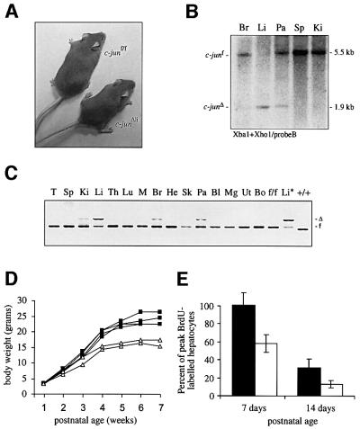
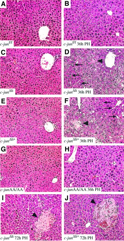
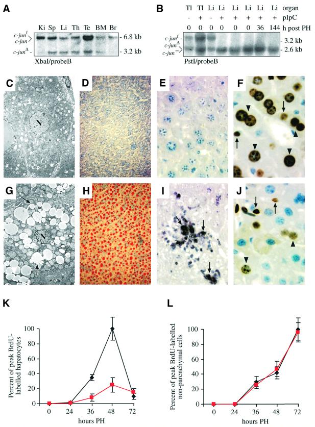
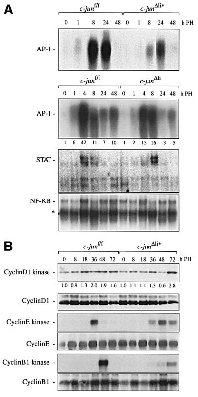
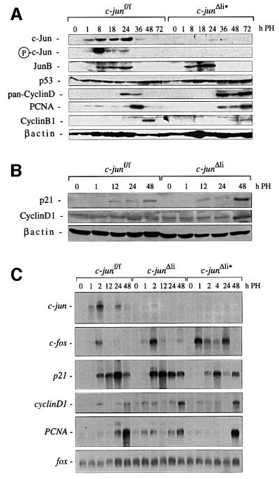
Mice lacking the AP-1 transcription factor c-jun die at mid-gestation showing heart defects and impaired hepatogenesis. To inactivate c-jun in hepatocytes, mice carrying a floxed c-jun allele were generated. Perinatal liver-specific c-jun deletion caused reduced hepatocyte proliferation and decreased body size. After partial hepatectomy, half of the mutants died and liver regeneration was impaired. This phenotype was not present in mice lacking the N-terminal phosphorylation sites of c-Jun. The failure to regenerate was accompanied by increased cell death and lipid accumulation in hepatocytes. Moreover, cyclin-dependent kinases and several cell cycle regulators were affected, resulting in inefficient G(1)-S phase progression. These studies identify c-Jun as a critical regulator of hepatocyte proliferation and survival during liver development and regeneration.


Figures

None
Fig. 1. Generation of mice harbouring a floxed c-jun allele. (A) Schematic representation of the targeting strategy employed to generate a floxed allele of c-jun. The c-jun open reading frame is represented by a rectangle, thin lines represent untranslated regions of the c-jun locus. The neomycine resistance gene (NeoR), the thymidine kinase gene (tk) and the diphtheria toxin alpha gene (DTα) are indicated; loxP sites are shown as triangles. X, XbaI; Xh, XhoI; P, PstI; E, EcoRI; H, HindIII. (B) Southern blot analysis of genomic DNA from the parental ES cell line E14.1 (+/+) and from three targeted (targ/+) ES cell clones. (C) After transient tranfection of cre, ES cell clones were selected for the loss of the NeoR and tk genes. Southern blot analysis of genomic DNA from the parental ES cell line E14.1 (+/+) and from three floxed (f/+) ES cell clones.
None
Fig. 2. Analysis of mice lacking c-jun in the liver. (A) Photograph of 8-week-old female c-junΔli and littermate control mice. (B) Southern blot of genomic DNA isolated from various organs of a c-junf/f Alfp-cre mouse (c-junΔli). Br, brain; Li, liver; Pa, pancreas; Sp, spleen; Ki, kidney. (C) PCR analysis of genomic DNA isolated from various organs of a c-junΔli mouse. T, tail; Sp, spleen; Ki, kidney; Li, liver; Th, thymus; Lu, lung; M, skeletal muscle; Br, brain, He, Heart; Sk, skin; Pa, pancreas; Bl, bladder; Mg, mammary gland; Ut, uterus; Bo, bone; f/f, c-junf/f control; Li*, liver of c-junf/f Mx-cre (c-junΔli*) mouse; +/+, wild-type control. (D) Postnatal growth rate of one litter of c-junf/f (black squares) and c-junΔli mice (white triangles). The result is representative of six litters analysed from three independent breeding cages. (E) Quantification of BrdU-immunostaining of c-junf/f (black bar) and c-junΔli livers (white bar) 7 and 14 days after birth.
None
Fig. 3. c-jun is required for liver regeneration. Histological analysis of wild-type (A, B), c-junΔli (Alfp-cre; C, D, I), c-junΔli* (Mx-cre; E, F, J) and junAA/AA (G, H) livers before (A, C, E, G), 36 h after (B, D, F, H) and 72 h after (I, J) hepatectomy. In (D), (F), (I) and (J), arrowheads indicate areas of necrotic tissue; some fat-containing vacuoles are indicated by arrows (magnification ×20).
None
Fig. 4. Increased cell death and decreased hepatocyte proliferation after hepatectomy in the absence of c-jun. (A) Southern blot analysis of genomic DNA isolated from various organs of one pIpC-treated c-junf/+ Mx-cre+ mouse. Ki, kidney; Sp, spleen; Li, liver; Th, thymus; Te, testes; BM, bone marrow; Br, brain. (B) Southern blot analysis of genomic DNA isolated from tail (Tl) and liver (Li) of pIpC-treated (+) and untreated (–) c-junf/– heterozygous mice before and after hepatectomy. c-jun is the null allele (Hilberg et al., 1993). (C, G) Electronmicroscope image of a control (C) and a c-junΔli mutant liver (G) 48 h after hepatectomy. The hepatocyte nucleus is indicated by ‘N’ and fat-containing vacuoles by arrows. (D, H) Oil red O-staining of a control (D) and a c-junΔli* mutant (H) liver 48 h after hepatectomy. Neutral lipids are visualized as red droplets in (H). (E, I) TUNEL-staining of a control (E) and a c-junΔli* mutant liver (I) 48 h after hepatectomy. Some TUNEL-positive cells are indicated by arrows. (F, J) BrdU-immunostaining of c-junf/f (F) and c-junΔli* (J) livers 48 h after hepatectomy. Arrows and arrowheads point to some BrdU-positive non- parenchymal cells and hepatocytes, respectively (J). (K, L) Quantification of BrdU-positive hepatocytes (K) and non-parenchymal cells (L) of c-junf/f (black lines) and c-junΔli* (red lines) mice after hepatectomy. Magnification: (C, G), ×3150; (D, H), ×20; (E, F, I, J), ×40.
None
Fig. 5. Molecular analysis of regenerating livers lacking c-jun. (A) AP-1, STAT and NF-κB DNA-binding in nuclear extracts from c-junf/f, c-junΔli and c-junΔli* livers; numbers below the AP-1 electrophoretic mobility-shift assay of c-junf/f and c-junΔli livers represent the fold induction of DNA-binding activity relative to the respective 0 time point; the asterisk indicates binding of p50 homodimers. (B) Immuno precipitated cyclin D1-, cyclin E- or cyclin B1-associated kinase activity in c-junf/f and c-junΔli* livers isolated at the indicated times after hepatectomy. Numbers below the cyclin D1-kinase activity represent the fold induction relative to the respective 0 time point. The presence of cyclin D1, cyclin E and cyclin B1 in the immunoprecipitates was confirmed by western blot analysis.
None
Fig. 6. Protein and RNA expression in regenerating livers lacking c-jun. (A, B) Expression of various proteins in c-junf/f, c-junΔli* (A) and c-junΔli (B) livers isolated at the indicated time points after hepatectomy. β-actin was used as a loading control. (C) Induction of c-jun, c-fos and fox mRNA (loading control), p21, PCNA and cyclinD1 in regenerating c-junf/f and c-jun-mutant livers at the indicated time points after hepatectomy.

References

    1. Angel P., Hattori,K., Smeal,T. and Karin,M. (1988) The jun proto-oncogene is positively autoregulated by its product, Jun/AP1. Cell, 55, 875–885. - PubMed
    1. Arias J., Alberts,A.S., Brindle,P., Claret,F.X., Smeal,T., Karin,M., Feramisco,J. and Montminy,M. (1994) Activation of cAMP and mitogen responsive genes relies on a common nuclear factor. Nature, 370, 226–229. - PubMed
    1. Bakiri L., Lallemand,D., Bossy-Wetzel,E. and Yaniv,M. (2000) Cell cycle-dependent variations in c-Jun and JunB phosphorylation: a role in the control of cyclin D1 expression. EMBO J., 19, 2056–2068. - PMC - PubMed
    1. Bannister A.J., Oehler,T., Wilhelm,D., Angel,P. and Kouzarides,T. (1995) Stimulation of c-Jun activity by CBP: c-Jun residues Ser63/73 are required for CBP induced stimulation in vivo and CBP binding in vitro. Oncogene, 11, 2509–2514. - PubMed
    1. Behrens A., Sibilia,M. and Wagner,E.F. (1999) Amino-terminal phosphorylation of c-Jun regulates stress-induced apoptosis and cellular proliferation. Nature Genet., 21, 326–329. - PubMed

Publication types

Substances