. 2002 Apr 1;157(1):125-36.
doi: 10.1083/jcb.200108089. Epub 2002 Apr 1.

Muscle-specific RING finger-1 interacts with titin to regulate sarcomeric M-line and thick filament structure and may have nuclear functions via its interaction with glucocorticoid modulatory element binding protein-1

Affiliations

Muscle-specific RING finger-1 interacts with titin to regulate sarcomeric M-line and thick filament structure and may have nuclear functions via its interaction with glucocorticoid modulatory element binding protein-1

Abigail S McElhinny et al. J Cell Biol. .

Abstract

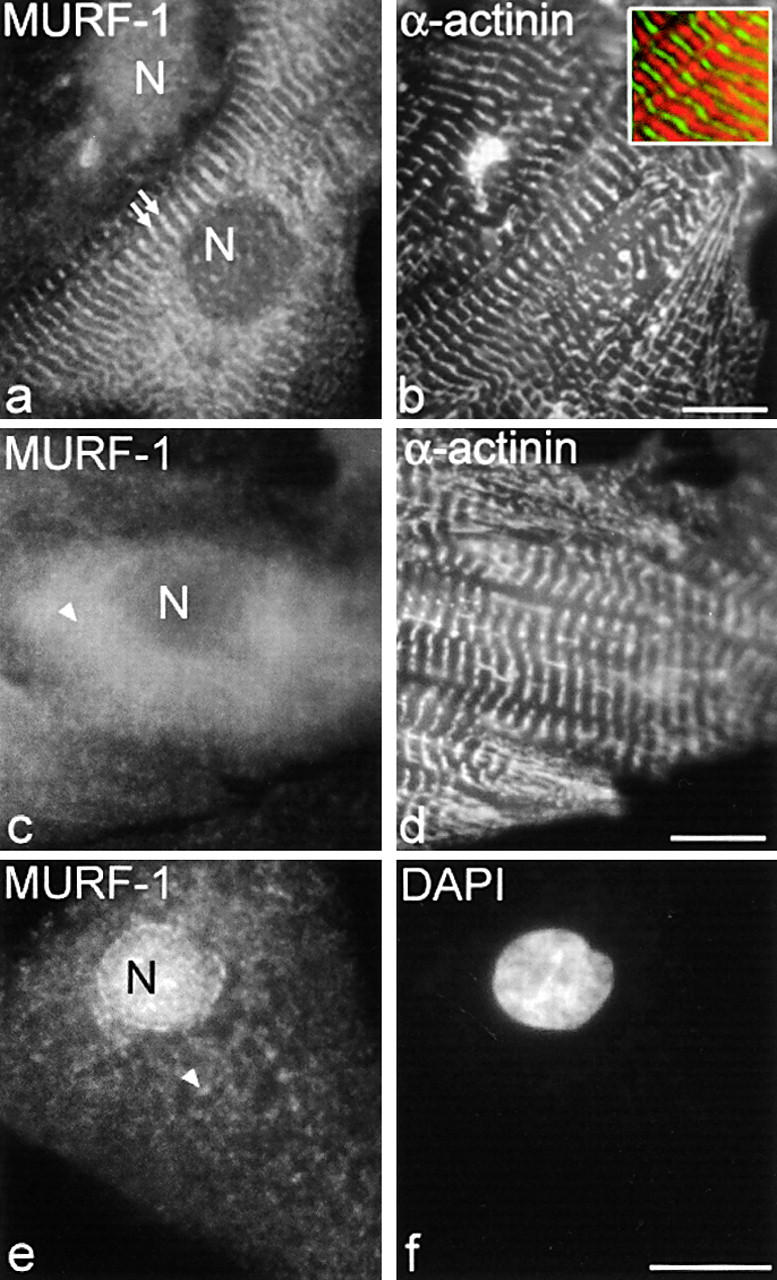
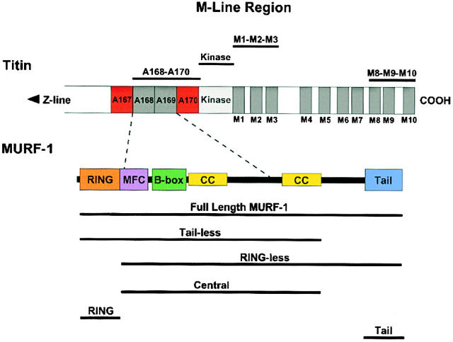
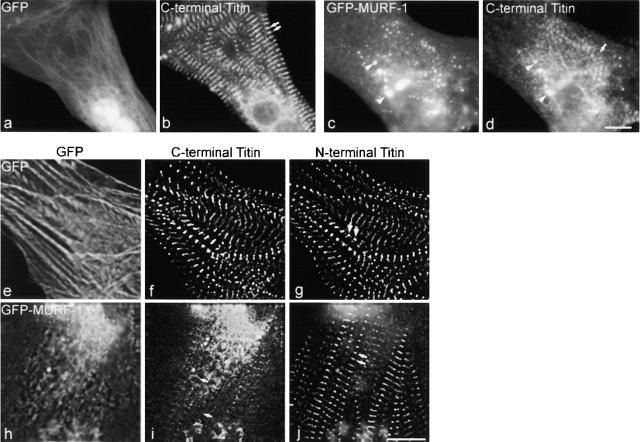
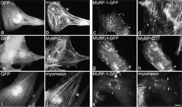
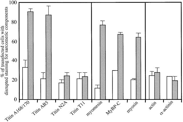
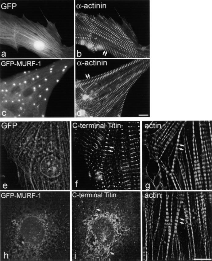
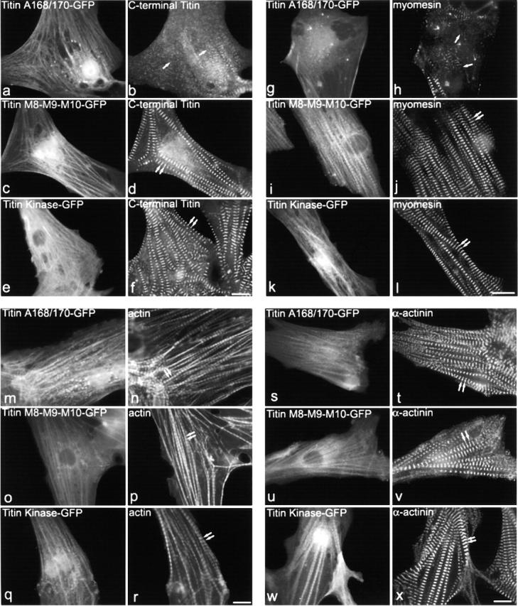
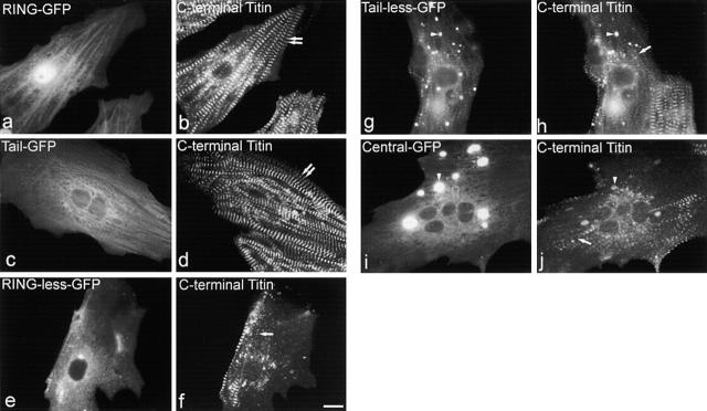
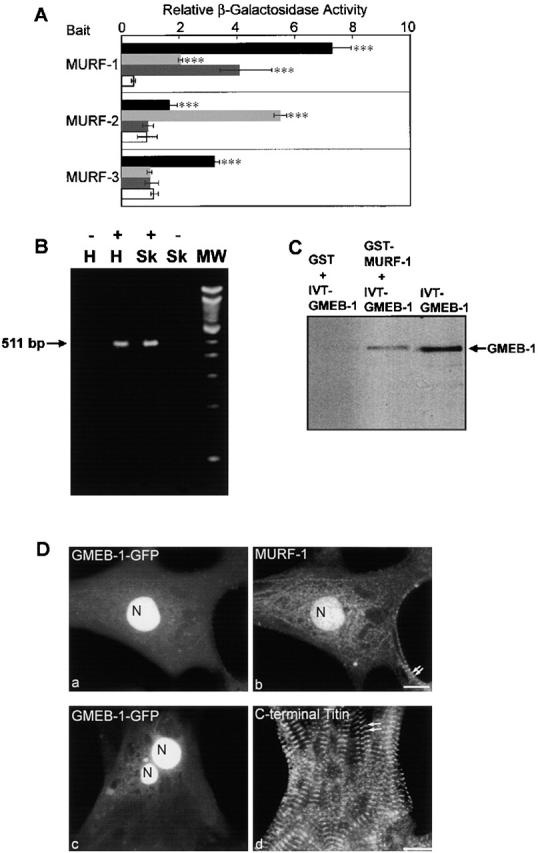
The COOH-terminal A168-170 region of the giant sarcomeric protein titin interacts with muscle-specific RING finger-1 (MURF-1). To investigate the functional significance of this interaction, we expressed green fluorescent protein fusion constructs encoding defined fragments of titin's M-line region and MURF-1 in cardiac myocytes. Upon expression of MURF-1 or its central region (containing its titin-binding site), the integrity of titin's M-line region was dramatically disrupted. Disruption of titin's M-line region also resulted in a perturbation of thick filament components, but, surprisingly, not of the NH2-terminal or I-band regions of titin, the Z-lines, or the thin filaments. This specific phenotype also was caused by the expression of titin A168-170. These data suggest that the interaction of titin with MURF-1 is important for the stability of the sarcomeric M-line region.MURF-1 also binds to ubiquitin-conjugating enzyme-9 and isopeptidase T-3, enzymes involved in small ubiquitin-related modifier-mediated nuclear import, and with glucocorticoid modulatory element binding protein-1 (GMEB-1), a transcriptional regulator. Consistent with our in vitro binding data implicating MURF-1 with nuclear functions, endogenous MURF-1 also was detected in the nuclei of some myocytes. The dual interactions of MURF-1 with titin and GMEB-1 may link myofibril signaling pathways (perhaps including titin's kinase domain) with muscle gene expression.


Figures

Figure 1.
Figure 1.
MURF-1 is detected in the M-line region of the sarcomere, diffuse in the cytoplasm, and in nuclei in fetal rat cardiac myocytes. Rat cardiac myocytes were labeled with polyclonal anti–MURF-1 antibodies followed by Texas red–conjugated secondary antibodies (a, c, and e), and with monoclonal α-actinin antibodies followed by Cy2-conjugated secondary antibodies (b and d). DAPI stain was added to identify nuclei (f). Note, inset in b is a merged image of MURF-1 (red) and α-actinin (green) staining. MURF-1 staining in the M-line region (a, double arrows), diffuse (c and e, arrowheads), and/or in nuclei with varying staining intensities (a and e). N, nucleus. Bars, 10 μm.
Figure 2.
Figure 2.
The COOH-terminal region of titin, revealing its Ig, FN III, and Ser/Thr kinase domains (modified from Centner et al., 2001). Most of the full-length titin molecule is composed of repeating Ig (dark gray) and FN III (red) modules, but also contains 17 unique domains, including a Ser/Thr kinase domain at the M-line region (light gray). The MURF-1 binding site resides within the Ig and FN domains A168–170, just NH2-terminal to the kinase domain. Bars above the titin molecule denote constructs used. Schematic structure of MURF-1 shown under titin, including its RING domain (orange), MURF family conserved region (purple), B-box domain (green), coiled-coil domains (yellow), and tail region (blue). The titin A168–170 binding site is located within the central region of MURF-1. The GFP–MURF-1 deletion constructs are shown under full-length MURF-1.
Figure 3.
Figure 3.
Expression of GFP–MURF-1 results in severe disruption of titin's M-line structure. Cells expressing GFP–MURF-1 (c) or GFP alone (a) were stained with anti-titin A168–170 antibodies followed by Texas red– conjugated secondary antibodies (b and d). In most myocytes, GFP–MURF-1 expression resulted in a severe disruption of the M-line region of titin (d) compared with myocytes expressing GFP alone (b). Note, titin staining colocalized with many of the GFP–MURF-1 aggregates (c and d, arrowheads). The disruption of titin appears to be localized to its COOH-terminal region. Triple-labeling studies in GFP- (e) and GFP–MURF-1–expressing (h) cells revealed that epitopes from the NH2-terminal region of titin (j) appeared regular and striated in the same myofibrils exhibiting disrupted COOH-terminal titin staining (i). Double arrows mark regular, striated titin and single arrows mark disrupted titin. Bars, 10 μm.
Figure 4.
Figure 4.
Expression of GFP–MURF-1 also perturbs the organization of thick filament components. Transfected myocytes were stained with antibodies to myosin (b and d), MyBP-C (f and h), and myomesin (j and l). In many GFP–MURF-1–expressing myocytes, staining for thick filament components was perturbed (d, h, and l), compared with myocytes transfected with GFP alone (b, f, and j). Single arrows mark perturbed thick filament component staining, and double arrows mark regular, striated thick filament staining. Note, size and intensity of the GFP–MURF-1 cytoplasmic aggregates vary from cell to cell (c, g, and k, arrowheads). Bar, 10 μm.
Figure 6.
Figure 6.
Quantification of GFP–MURF-1– and GFP-expressing myocytes exhibiting disrupted staining patterns for sarcomeric components. Myocytes expressing GFP–MURF-1 (gray bars) or GFP alone (white bars) were stained for various sarcomeric components, and the number of cells exhibiting perturbed staining patterns was counted. The results indicate that the COOH-terminal region of titin (A168–170 and AB5) was severely disrupted in most GFP–MURF-1–expressing cells, whereas its I-band (N2A) and NH2-terminal region (T11) were not affected. The thick filament components myosin, myomesin, and C-protein were also disrupted in a large majority of GFP–MURF-1–expressing cells, but thin filament and Z-line components appear relatively unaffected. Data are presented as the mean percentage of total myocytes with disrupted staining ± SD. Means were obtained by counting >50 myocytes from more than two experiments and the results are representative of >10 experiments.
Figure 5.
Figure 5.
Expression of GFP–MURF-1 does not appear to affect the integrity of thin filament or Z-line components. Myocytes expressing GFP–MURF-1 (c) or GFP alone (a) were stained for Z-lines with saromeric α-actinin antibodies (b and d) and show regular, striated staining. Triple-labeling studies in GFP–MURF-1–transfected cells (h), using Texas red–conjugated phalloidin (j) and antibodies to titin A168–170 (i), determined that thin filament integrity is not affected upon disruption of COOH-terminal titin in identical myofibrils. GFP-transfected cells (e) exhibited normal actin filament (g) and COOH-terminal titin (f) staining. Double arrows mark regular, striated staining. Single arrows mark disrupted titin staining. Bars, 10 μm.
Figure 7.
Figure 7.
Expression of titin domains A168–170 also disrupts titin M-line region and thick filament structure, but not thin filaments or Z-lines. Myocytes expressing titin A168–170 or other Ig domains from the M-line region of titin were stained for various sarcomeric components. Costaining of A168-170–GFP expressing myocytes with anti– C-terminal titin region antibodies revealed a severe disruption of titin (a and b), compared with myocytes transfected with M8-M9-M10-GFP (c and d) or titin kinase–GFP (e and f). Costaining of titin A168-170–transfected myocytes with antibodies against thick filament components, including myomesin (g and h), reveals that the integrity of the thick filaments is perturbed compared with myocytes transfected with titin M8-M9-M10-GFP (i and j) or titin kinase–GFP (k and l). Myocytes transfected with titin A168-170–GFP (m, n, s, and t), titin M8-M9-M10–GFP (o, p, u, and v), or titin kinase–GFP (q, r, w, and x) were stained with Texas red–conjugated phalloidin (n, p, and r) or antibodies to sarcomeric α-actinin (t, v, and x). Double arrows mark regular, striated staining. Single arrows mark disrupted staining. Bars, 10 μm.
Figure 8.
Figure 8.
The central portion of MURF-1 is involved in maintaining the structure of COOH-terminal titin. Myocytes expressing defined regions of MURF-1 were stained with anti-titin A168–170 antibodies. Expression of the RING–GFP (a and b) or Tail–GFP (c and d) proteins did not appear to affect the integrity of COOH-terminal titin. Expression of GFP fusion proteins of the central MURF region (Central; i and j), the RING plus central regions (Tailless; g and h), and the central plus tail regions (RINGless; e and f) severely disrupted titin's COOH-terminal region (single arrow marks disrupted titin A168–170 staining; double arrows mark regular, striated titin A168–170 staining). Note that titan staining colocalized with many of the GFP–MURF-1 aggregates (g–j, arrowheads). Bar, 10 μm.
Figure 9.
Figure 9.
MURF family members interact with SUMO modifying enzymes ISOT-3 and Ubc9, but only MURF-1 interacts with the transcriptional regulator GMEB-1. (A) Y2H screens using full-length cDNAs of individual MURF family members as baits identified ISOT-3 (light gray) and Ubc9 (black) as MURF-binding proteins. However, GMEB-1 (dark gray) was found to interact only with MURF-1. β-Galactosidase assays were performed to confirm positive clones, and the levels were compared with colonies transformed with each prey construct and the empty bait vector (white). Data are presented as mean levels of β-galactosidase from triplicate experiments ± SD. ***, P > 0.001. (B) RT-PCR analysis of human heart total RNA revealed that GMEB-1 mRNA transcripts are detectable in heart (H) and skeletal (Sk) tissues. Lane 1, no reverse transcriptase control in human heart RNA (−); lane 2, 511-bp GMEB-1 PCR product amplified from human heart RNA (+); lane 3, 511-bp GMEB-1 PCR product amplified from human skeletal RNA (+); lane 4, no reverse transcriptase control in human skeletal RNA (−). (C) GMEB-1 specifically binds to MURF-1 in GST pull-down assays. GMEB-1 was translated in vitro (lane 3). When incubated with bacterially expressed GST–MURF-1 fusion peptides, GMEB-1 and MURF-1 binding to glutathione–sepharose 4B beads was detectable (lane 2). Lane 1 contains no detectable binding of GMEB-1 to the beads alone. IVT, in vitro translated. (D) GMEB-1–GFP targets to the nuclei of cardiac myocytes (a and c). MURF-1 staining also was present in some of the nuclei that contained GMEB-1–GFP (b). Note, MURF-1 is also detected at the M-line region in the same myocytes (b, double arrows). Expression of GMEB-1–GFP in cardiac myocytes does not appear to affect the integrity of the COOH-terminal region of titin (d, staining with anti-titin A168–170 antibodies). Double arrows mark regular, striated titin staining. N, nuclei. Bars, 10 μm.

References

    1. Antin, P.B., S. Forry-Schaudies, T.M. Friedman, S.J. Tapscott, and H. Holtzer. 1981. Taxol induces postmitotic myoblasts to assemble interdigitating microtubule-myosin arrays that exclude actin filaments. J. Cell Biol. 90:300–308. - PMC - PubMed
    1. Bang, M.L., T. Centner, F. Fornoff, A.J. Geach, M. Gotthardt, M. McNabb, C.C. Witt, D. Labeit, C.C. Gregorio, H. Granzier, and S. Labeit. 2001. The complete gene sequence of titin, expression of an unusual approximately 700-kDa titin isoform, and its interaction with obscurin identify a novel Z-line to I-band linking system. Circ. Res. 89:1065–1072. - PubMed
    1. Bodine, S.C., E. Latres, S. Baumhuerer, V.K. Lai, L. Nunez, B.A. Clarke, W.T. Poueymirou, F.J. Panaro, E. Na, K. Dharmarajan, Z.Q. Pan, D.M. Valenzuela, T.M. DeChiara, T.N. Stirr, G.D. Yancopoulos, and D.J. Glass. 2001. Identification of ubiquitin ligases required for skeletal muscle atrophy. Science. 294:1704–1708. - PubMed
    1. Borden, K.L. 2000. RING domains: master builders of molecular scaffolds? J. Mol. Biol. 295:1103–1112. - PubMed
    1. Centner, T., F. Fougerousse, A. Freiburg, C. Witt, J.S. Beckmann, H. Granzier, K. Trombitas, C.C. Gregorio, and S. Labeit. 2000. Molecular tools for the study of titin's differential expression. Adv. Exp. Med. Biol. 481:35–49. - PubMed

Publication types

MeSH terms

Associated data