. 2002 Apr 1;540(Pt 1):57-72.
doi: 10.1113/jphysiol.2001.013294.

Control of the propagation of dendritic low-threshold Ca(2+) spikes in Purkinje cells from rat cerebellar slice cultures

Affiliations

Control of the propagation of dendritic low-threshold Ca(2+) spikes in Purkinje cells from rat cerebellar slice cultures

Pauline Cavelier et al. J Physiol. .

Abstract

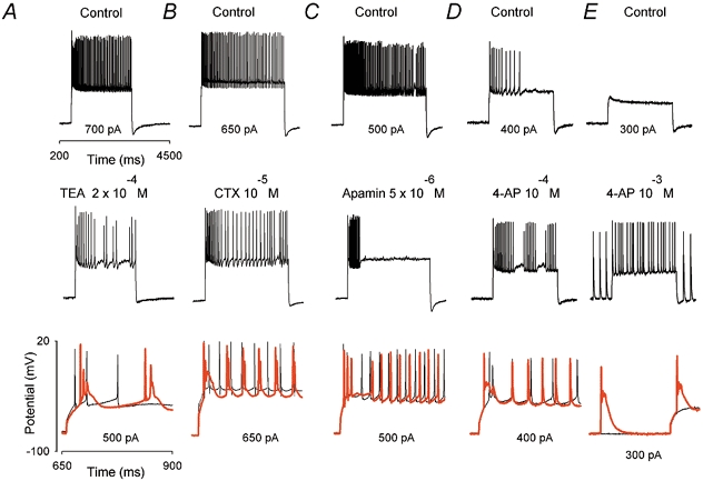
To investigate the ionic mechanisms controlling the dendrosomatic propagation of low-threshold Ca(2+) spikes (LTS) in Purkinje cells (PCs), somatically evoked discharges of action potentials (APs) were recorded under current-clamp conditions. The whole-cell configuration of the patch-clamp method was used in PCs from rat cerebellar slice cultures. Full blockade of the P/Q-type Ca(2+) current revealed slow but transient depolarizations associated with bursts of fast Na(+) APs. These can occur as a single isolated event at the onset of current injection, or repetitively (i.e. a slow complex burst). The initial transient depolarization was identified as an LTS Blockade of P/Q-type Ca(2+) channels increased the likelihood of recording Ca(2+) spikes at the soma by promoting dendrosomatic propagation. Slow rhythmic depolarizations shared several properties with the LTS (kinetics, activation/inactivation, calcium dependency and dendritic origin), suggesting that they correspond to repetitively activated dendritic LTS, which reach the soma when P/Q channels are blocked. Somatic LTS and slow complex burst activity were also induced by K(+) channel blockers such as TEA (2.5 x 10(-4) M) charybdotoxin (CTX, 10(-5) M), rIberiotoxin (10(-7) M), and 4-aminopyridine (4-AP, 10(-3) M), but not by apamin (10(-4) M). In the presence of 4-AP, slow complex burst activity occurred even at hyperpolarized potentials (-80 mV). In conclusion, we suggest that the propagation of dendritic LTS is controlled directly by 4-AP-sensitive K(+) channels, and indirectly modulated by activation of calcium-activated K(+) (BK) channels via P/Q-mediated Ca(2+) entry. The slow complex burst resembles strikingly the complex spike elicited by climbing fibre stimulation, and we therefore propose, as a hypothesis, that dendrosomatic propagation of the LTS could underlie the complex spike.


Figures

Figure 1
Figure 1. Somatically evoked responses recorded after the blockade of P/Q-type Ca2+ channels reveal slow but transient depolarizations
A-C, responses to current injections recorded after preincubation with toxins that block P/Q-type Ca2+ channels. A, an initial transient depolarization (shown on an expanded time scale in the inset, with an amplitude and duration as indicated by arrows) triggering a fast action potential (AP). This initial response is followed by a depolarizing plateau. B, an initial slow, transient depolarization triggering a fast AP, which is followed by slow depolarizing waves, each of which elicited fast APs. C, a slow complex burst with amplitude and duration as indicated by arrows. D, frequency of slow complex bursts (typified in C) as a function of injected current intensity. E, time course of the effects on the evoked discharge of a drop of ω-agatoxin TK (2 × 10⁻⁷m) applied close to the recorded cell. F, effects on the evoked discharge of Cd2+ (5 × 10⁻⁵m). In both cases, the left traces are the control responses (regular firing of fast APs) and the middle traces are the responses after the application of either toxin or Cd2+ (a firing of slow complex bursts is induced as shown by the third traces on an expanded time scale). The right panel illustrates the initial response under control conditions (thin trace) and after toxin and Cd2+ application (red thick trace). Note the appearance of an initial slow but transient depolarization in the presence of toxin or Cd2+.
Figure 2
Figure 2. The initial transient depolarization recorded in the soma after blockade of P/Q-type Ca2+ channels is a Ca2+ AP with a low activation threshold
A, in all cases the time scale is shown between 200 and 500 ms to illustrate the initial response. The initial transient depolarization is insensitive to TTX (first panel from top). The left trace is the control response and the right trace is the response after TTX (5 × 10⁻⁷m). The initial transient depolarization is not sodium-dependent (second panel from top). The left trace shows the evoked response recorded in the presence of TTX and the right trace the response after perfusion of a sodium-free external solution. The initial transient slow depolarization is calcium-dependent (third panel from top). The left trace shows the initial transient depolarization isolated in the presence of TTX, and the right trace the response after perfusion with a calcium-free solution. B, activation of the calcium-dependent initial response isolated in the presence of TTX. Responses evoked with current injections of increasing intensity are shown. The threshold potential for the transient depolarization is indicated by the arrow: during the current injection the cell first depolarized, following an exponential time course (thick line) until (arrow) the slow AP was evoked. C, the probability of recording a Ca2+ spike depends upon the recording site. Histogram of the percentage of recordings displaying a Ca2+ spike as a function of the distance along the dendrite under control conditions (black bars) and after preincubation with ω-agatoxin TK or ω-conotoxin MVII C to block P/Q channels (white bars).
Figure 6
Figure 6. Effects of apamin and 4-AP on evoked responses recorded after the blockade of P/Q-type Ca2+ channels
To reveal slow complex burst firing, cultured slices were preincubated with ω-agatoxin-TK for at least 15 min to block P/Q-type Ca2+ channels. A, effects of apamin. The left trace is the control response; the middle trace and the right trace represent the response 50 s and 75 s after apamin treatment, respectively. Note that the repetitive slow complex bursts were abolished by apamin after 75 s. The left panel illustrates the control response on an expanded time scale (thin black trace) and the response during application of apamin (thick red trace), to show that apamin reduced the slow hyperpolarization that followed the slow complex burst. B, C, effects of 4-AP (10−3m). B, the left trace is the control response (only an initial low-threshold Ca2+ spike was evoked) and the middle trace and the right trace are responses 35 s and 45 s after 4-AP treatment, respectively. The right panel illustrates the control response on an expanded time scale (black thin trace) and the response during application of 4-AP (red thick trace). Note the appearance of a discharge of slow complex bursts. C, the left trace is the control response (a discharge of slow complex bursts), the middle trace is the response after bath application of TTX (5 × 10⁻⁷m; only an initial low-threshold Ca2+ spike is evoked), and the right trace is the response after a bath application of 4-AP (10−3m) in the presence of TTX (a discharge of low-threshold Ca2+ spikes was induced).
Figure 3
Figure 3. Comparison of the effect of saccharose application on the soma and on the main dendrite reveals a dendritic initiation site for the low-threshold Ca2+ spike with or without functional P/Q-type Ca2+ channels
To the left of the figure are schematic diagrams of the protocol indicating that the recording pipette was on the soma and a second pipette was used to apply saccharose (dashed lines) onto either the soma (second drawing from top) or the main dendrite (fourth drawing from top). A and B, recordings from the same cell illustrating, under control conditions, sustained discharge of fast APs (see the traces on top) in response to a depolarizing current injection. The lower traces in A and B illustrate the response after application of saccharose onto the soma and onto the dendrite, respectively. The repetitive response of fast APs was either totally or partially abolished following the somatic or dendritic location of the saccharose application. C and D, recordings from another cell. Under control conditions, the evoked response was an initial low-threshold Ca2+ spike with one fast AP on top (see the traces on top). The lower traces in C and D illustrate the response after application of saccharose onto the soma and onto the dendrite, respectively. The low-threshold Ca2+ spike was affected only when saccharose was applied onto the main dendrite. E and F, the same cell recorded after incubation with a toxin that blocks P/Q-type Ca2+ channels. The top traces in E and F illustrate the control responses to a depolarizing current injection (an initial low-threshold Ca2+ spike with a fast AP on top). The lower traces in E and F illustrate the response after application of saccharose onto the soma and onto the dendrite, respectively. Again, the initial low-threshold Ca2+ spike was only affected when saccharose was perfused onto the dendrite.
Figure 4
Figure 4. Activation-inactivation properties and ionic dependence of the slow complex burst discharge recorded after preincubation with toxins against P/Q channels
A, complex burst firing recorded at different holding potentials (left panel) and the amplitude of the slow depolarizing wave (measured as indicated in the inset) as a function of the holding potential (right panel). B, ionic dependence of the slow complex burst discharge. The upper traces illustrate control responses. The lower trace on the left is obtained after perfusion with calcium-free external solution. Only a fast AP is elicited at the onset of the step. The lower trace in the middle represents the response obtained after bath application of TTX (5 × 10⁻⁷m): in this condition only a slow AP was triggered at the beginning of the step. The lower trace on the right shows the response recorded in sodium-free external solution, where only a slow AP is elicited at the beginning of the step. C, Somatic and dendritic conductances were required for a repetitive activation of the slow complex bursts revealed after the blockade of P/Q-type Ca2+ channels. The control response (protocol illustrated by the first drawing) recorded after incubation with ω-agatoxin-TK was a slow complex burst (upper trace on the left) and is shown on an expanded time scale (lower trace). The middle and right traces illustrate the responses after perfusion of saccharose onto the soma and onto the dendrite, respectively. The lower traces illustrate these responses on an expanded time scale. Saccharose applied onto the soma abolished repetitive firing, and saccharose applied onto the main dendrite after recovery to the control response (not shown) only blocked slow spikes.
Figure 5
Figure 5. Effects of K+ channels blockers on the firing of fast APs: BK channel blockers and 4-aminopyridine (4-AP), but not apamin, induced firing of slow complex bursts and the appearance of an initial Ca2+ spike
A-E, effects of TEA (2 × 10⁻⁴m), charybdotoxin (CTX; 10−5m), apamin (10−4m), 4-AP (10−4m) and 4-AP (10−3m) on the evoked discharge, respectively. Upper traces are control responses and middle traces are responses recorded after bath application of K+ channel blockers. The lower traces illustrate the initial response on an expanded time scale under control conditions (thin trace) and after the application of a K+ channel blocker (red thick trace). TEA and CTX induced the appearance of an initial low-threshold Ca2+ spike and slow complex burst. Apamin reduced the duration of fast AP discharge. 4-AP applied at 10−4m induced an initial low-threshold Ca2+ spike followed by fast APs, whereas application of 4-AP at 10−3m induced slow complex bursts, even at −80 mV.
Figure 7
Figure 7. Spontaneous activity of Purkinje cells (PCs): simple spikes and slow complex bursts
Spontaneous activity of PCs recorded at the resting membrane potential. A, sustained regular discharge of fast APs is recorded. B, regular discharge of slow complex bursts. C, simple spikes and slow complex bursts evoked on depolarizing plateaus.
Figure 8
Figure 8. Evoked responses of PCs recorded from acute slices. Co-application of TEA and Cd2+ induced firing of slow complex bursts
A, evidence for an initial slow depolarization that is insensitive to TTX. The evoked response to current injection is illustrated on an expanded time scale to show the initial part of the response. The thin black trace illustrates an initial slow depolarization triggering a burst of fast APs, and the red thick trace shows the response after TTX, where an initial slow transient depolarization was isolated. B, D, effects of TEA (2.5 × 10⁻⁴m) and Cd2+ (5 × 10⁻⁵m) on the evoked discharge. B, the left trace is the control response (sustained firing of fast APs), the middle trace is the response after TEA (burst of fast APs), and the right trace shows the response when Cd2+ was applied in the presence of TEA, revealing slow complex bursts. C, the initial response under control conditions on an expanded time scale (black thin trace) and after a co-application of TEA and Cd2+ (red thick trace) to show that TEA + Cd2+ reveals an initial low-threshold Ca2+ spike. D, slow complex bursts on an expanded time scale, revealed by co-application of TEA and Cd2+ in a PC recorded from an acute slice (upper panel, same cell as in B) and from a slice culture (lower panel).

References

    1. Candia S, Garcia ML, Latorre R. Mode of action of iberiotoxin, a potent blocker of the large conductance Ca2+-activated K+ channel. Biophysical Journal. 1992;63:583–590. - PMC - PubMed
    1. Cerminara NL, Rawson JA. Distribution of calcium-activated potassium channels in the cerebellum of the rat. Society of Neurosciences Abstracts. 2000;255.6:73.
    1. Coetzee WA, Amarillo Y, Chiu J, Chow A, Lau D, McCormack T, Moreno H, Nadal MS, Ozaita A, Pountney D, Saganich M, Vega-Saenz de Miera E, Rudy B. Molecular diversity of K+ channels. Annals of the New York Academy of Sciences. 1999;29:233–285. - PubMed
    1. Colbert CM, Pan E. Arachidonic acid reciprocally alters the availability of transient and sustained dendritic K+ channels in hippocampal CA1 pyramidal neurons. Journal of Neurosciences. 1999;19:8163–8171. - PMC - PubMed
    1. Crepel F, Penit-Soria J. Inward rectification and low-threshold calcium conductance in rat cerebellar Purkinje cells. Journal of Physiology. 1986;372:1–23. - PMC - PubMed

Publication types

MeSH terms

LinkOut - more resources