Correlations between neuronal morphology and electrophysiological features in the rodent superficial dorsal horn
- PMID: 11927679
- PMCID: PMC2290200
- DOI: 10.1113/jphysiol.2001.012890
Correlations between neuronal morphology and electrophysiological features in the rodent superficial dorsal horn
Abstract
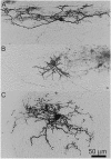
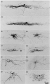
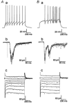
Relationships between the morphology of individual neurones of the spinal superficial dorsal horn (SDH), laminae I and II, and their electrophysiological properties were studied in spinal cord slices prepared from anaesthetized, free-ranging hamsters. Tight-seal, whole-cell recordings were made with pipette microelectrodes filled with biocytin to establish electrophysiological characteristics and to label the studied neurones. Neurones were categorized according to location and size of the somata, the dendritic and axonal pattern of arborization, spontaneous synaptic potentials, evoked postsynaptic currents, pattern of discharge to depolarizing pulses and current-voltage relationships. Data were obtained for 170 neurones; 13 of these had somata in lamina I and 157 in lamina II. Stimulation of the segmental dorsal root evoked a prompt excitatory response in almost every neurone sampled (161/166) with nearly 3/4 displaying putative monosynaptic EPSCs. The majority of neurones (133/170) fitted one of several distinctive morphological categories. To a considerable extent, neurones with a common morphological configuration and neurite disposition shared electrophysiological characteristics. Five of the 13 lamina I neurones were relatively large with extensive dendritic arborization in the horizontal dimension and a prominent axon directed ventrally and contralaterally. These presumptive ventrolateral projection neurones differed structurally and electrophysiologically from the other lamina I neurones, which had ipsilateral, locally arborizing axons and/or branches entering the dorsal lateral funiculus. One hundred and twenty lamina II neurones fitted one of five morphological categories: islet, central, medial-lateral, radial or vertical. Central cells were further divided into three groups on functional features. We conclude that the spinal SDH comprises many types of neurones whose morphological characteristics are associated with specific functional features implying diversity in functional organization of the SDH and in its role as a major synaptic termination for thin primary afferent fibres.
Figures


References
-
- Andrew D, Craig AD. Spinothalamic lamina I neurons selectively sensitive to histamine: a central neural pathway for itch. Nature Neuroscience. 2001;4:72–77. - PubMed
-
- Bao J, Li J, Perl ER. Primary afferent inhibition of spinal laminae I and II neurons. Society for Neuroscience Abstracts. 1996;22:878.
Publication types
MeSH terms
Substances
Grants and funding
LinkOut - more resources
Full Text Sources

