. 2002 Apr 2;99(7):4319-24.
doi: 10.1073/pnas.261702698.

FKBP12-rapamycin-associated protein associates with mitochondria and senses osmotic stress via mitochondrial dysfunction

Affiliations

FKBP12-rapamycin-associated protein associates with mitochondria and senses osmotic stress via mitochondrial dysfunction

Bimal N Desai et al. Proc Natl Acad Sci U S A. .

Abstract

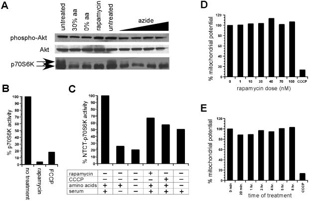
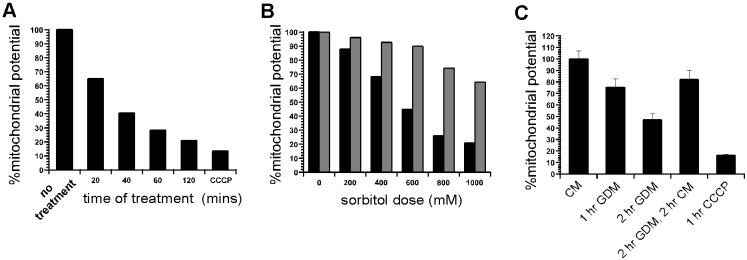
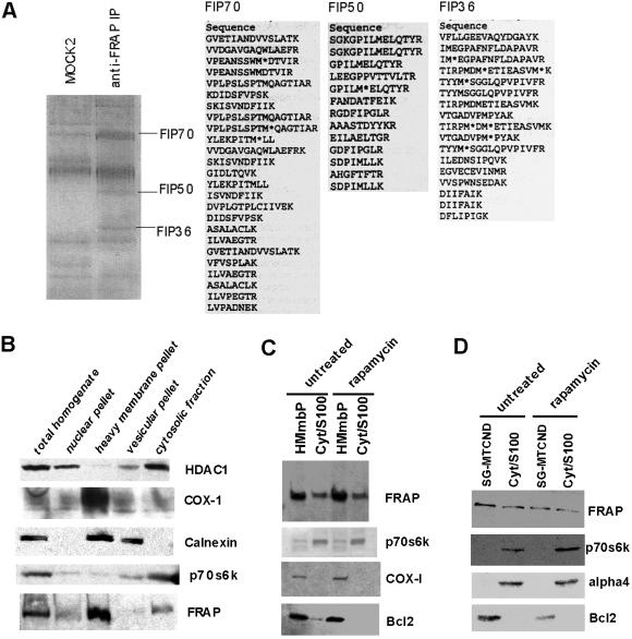
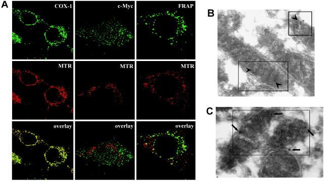
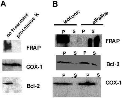
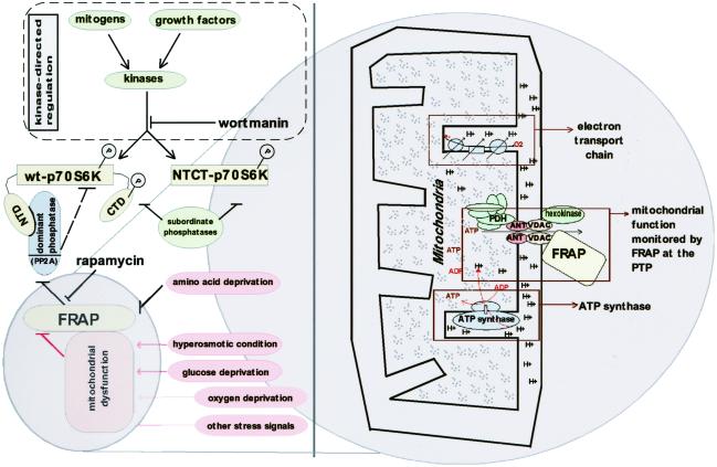
FKBP12-rapamycin associated protein (FRAP, also known as mTOR or RAFT) is the founding member of the phosphatidylinositol kinase-related kinase family and functions as a sensor of physiological signals that regulate cell growth. Signals integrated by FRAP include nutrients, cAMP levels, and osmotic stress, and cellular processes affected by FRAP include transcription, translation, and autophagy. The mechanisms underlying the integration of such diverse signals by FRAP are largely unknown. Recently, FRAP has been reported to be regulated by mitochondrial dysfunction and depletion of ATP levels. Here we show that exposure of cells to hyperosmotic conditions (and to glucose-deficient growth medium) results in rapid and reversible dissipation of the mitochondrial proton gradient. These results suggest that the ability of FRAP to mediate osmotic stress response (and glucose deprivation response) is by means of an intermediate mitochondrial dysfunction. We also show that in addition to cytosolic FRAP a large portion of FRAP associates with the mitochondrial outer membrane. The results support the existence of a stress-sensing module consisting of mitochondria and mitochondrial outer membrane-associated FRAP. This module allows the cell to integrate a variety of stress signals that affect mitochondrial function and regulate a growth checkpoint involving p70 S6 kinase.


Figures

Figure 1
Figure 1
Analysis of p70S6K phosphorylation in response to mitochondrial dysfunction. (A) p70S6K phospho-shift in response to untreated, 30% amino acid availability (30% aa), absence of amino acids (0% aa), rapamycin treatment, and increasing azide dosage (1 mM, 2 mM, 5 mM, 10 mM) is shown. Similar analysis of phospho-Akt and Akt is also included. (B) p70S6K activity in response to rapamycin and carbonylcyanide p-trifluoro-methoxyphenylhydrazone (FCCP) treatment. (C) Activity of ΔNTΔCT-p70S6K rapamycin-resistant allele in response to the presence of amino acids and serum, withdrawal of serum, withdrawal of amino acid and serum, rapamycin treatment, and carbonylcyanide m-chlorophenlyhydrazone (CCCP) treatment. (D) Mitochondrial activity in response to rapamycin dose is compared with CCCP-treated control measurement by using the JC-1 assay. (E) Mitochondrial activity in response to time of treatment with 50 nM rapamycin is compared with CCCP-treated control measurement.
Figure 2
Figure 2
Measurement of mitochondrial function in response to hyperosmotic conditions and glucose deprivation. (A) Mitochondrial activity in response to time of exposure to osmotic stress (600 mM sorbitol treatment) is compared with CCCP treatment. (B) Mitochondrial activity in response to level of osmotic stress (% sorbitol) is shown (black bars). Kit-225 cells were treated with sorbitol for 30 min before measuring mitochondrial activity. Mitochondrial activity 20 min after removal of osmotic stress (withdrawal of sorbitol) is shown as adjoining bars for each dose (gray bars). (C) Mitochondrial activity in response to glucose deprivation. Kit-225 cells were exposed to conditions indicated and mitochondrial proton gradient was measured by JC-1 assay. CM, complete media; GDM, glucose-deprived media.
Figure 3
Figure 3
Analysis of FRAP immune-complex and localization of FRAP by subcellular fractionation studies. (A) Identification of components of PDH complex in FRAP immune-complexes. Jurkat cells were lysed as described in Materials and Methods and the cell lysate was used to isolate FRAP immune-complexes by using an anti-FRAP polyclonal antibody. Coomassie-stained gel shows resolution of immmune-complex obtained by using a mock antibody (MOCK2) and the FRAP immune-complex (anti-FRAP IP). Three bands enriched in FRAP immune-complex but absent in mock preparations are marked according to their molecular weights. The number denotes the approximate molecular weight of the band. The bands labeled as FIP70, FIP50, and FIP36 were microsequenced to yield the peptide sequences shown for each band. (B) Equal amounts of indicated fractions were resolved by using SDS/PAGE and immuno-blotted for FRAP and marker proteins. Absence of HDAC1 and p70S6K in the heavy membrane pellet indicates that this fraction was not contaminated by significant amounts of nuclear or cytosolic proteins. Calnexin is predominantly an endoplasmic reticulum (ER)-resident protein; its presence in the heavy membrane pellet and the vesicular fraction is consistent with the fact that heavy membrane pellet contains mitochondrial, lysosomal, and ER-resident proteins. COX-1 is a mitochondrial marker and is therefore seen predominantly in the heavy membrane pellet. FRAP is present in heavy membrane pellet and cytosolic fraction. (C) Heavy membrane pellet (HMmbP) and cytosolic (Cyt/S-100) fractions from untreated and rapamycin-treated cells were analyzed for changes in the levels of FRAP and marker proteins. An additional mitochondrial marker (Bcl-2) is included in the analysis. (D) Mitochondrial fraction purified by using a sucrose gradient (SG-MTCND) and cytosolic fraction from untreated and rapamycin-treated cells was analyzed for the presence of FRAP.
Figure 4
Figure 4
Immunofluorescence-confocal microscopy and immuno-electron microscopy of FRAP in 3T3 cells. (A) Confocal microscopy shows colocolization of FRAP and mitochondria. MTR was used to stain the mitochondria before fixation and permeabilization. COX-1 was primarily stained with an anti-COX-1 mAb, FRAP was primarily stained with an anti-FRAP mAb, and c-myc was primarily stained with an anti-c-Myc mAb. An [Alexa-fluor-488]-conjugated antibody was used as the secondary stain. The yellow speckles in the overlay frames indicate colocolization. COX-1 shows complete colocolization (positive control for methodology) whereas c-Myc does not colocalize with mitochondria (negative control for methodology). A significant portion of FRAP is seen to colocalize with the mitochondria. (B and C) Immuno-electron microscopy indicates mitochondrial locolization of FRAP. A secondary stain of streptavidin-gold was used to detect the primary anti-FRAP mAb. Arrows point toward the electron-dense secondary probe within the boxes. In C the long arrows point toward typical membrane-proximal localizations that are seen at a high frequency. (Magnifications: ×15,000.)
Figure 5
Figure 5
Sensitivity of FRAPm to protease treatment and alkaline extraction. (A) Mitochondrial preparation with or without proteinase K treatment (20 min, room temperature) is resolved and immuno-blotted for FRAP and mitochondrial markers (COX-1 and Bcl-2). COX-1 is associated with the inner membrane and is therefore insensitive to proteinase K treatment. Bcl-2 is associated with the outer membrane and exposed, therefore sensitive to protease treatment. FRAP is also sensitive to protease treatment. (B) Analysis of isotonic and alkaline extraction of mitochondrial preparation is shown. The insoluble pellet (P) and the supernatant (S) of these extractions were resolved and probed for FRAP and marker proteins.
Figure 6
Figure 6
A model illustrating the role of FRAP in sensing mitochondrial dysfunction and regulation of p70S6K. The model highlights the role of FRAP in sensing signals initiated by hyperosmotic and glucose-deprivation conditions by an intermediate mitochondrial dysfunction. Other stress signals such as oxygen deprivation that result in mitochondrial dysfunction may also signal to FRAP by using similar mechanisms. FRAP senses amino acid deprivation by means of a mechanism independent of mitochondria. Two distinct pathways orchestrate the regulation of p70S6K: (i) the kinase-directed pathway initiated by mitogens or growth factors and in equilibrium with the “subordinate phosphatases” and (ii) the FRAP-mediated pathway, which involves a “dominant phosphatase” (a PP2A isoform) that is constitutively associated with wt-p70S6K by the N-terminal domain (NTD) of p70S6K. A rapamycin-resistant allele of p70S6K called NTCT-p70S6K is truncated at the NTD and the C-terminal domain (CTD). The dominant phosphatase is unable to associate with NTCT-p70S6K and therefore FRAP-mediated signals do not affect NTCT-p70S6K. A potential mechanism by which FRAP senses mitochondrial function is shown as a “zoom in” (Right). FRAP is shown to be associated with the mitochondrial PTP, which is composed of VDAC and ANT. The PDH complex (located in mitochondrial matrix) is shown in close proximity to PTP where it can receive glycolysis-derived pyruvate, which enters the mitochondria primarily by means of the PTP.

References

    1. Schmelzle T, Hall M N. Cell. 2000;103:253–262. - PubMed
    1. Brown E J, Beal P A, Keith C T, Chen J, Shin T B, Schreiber S L. Nature (London) 1995;377:441–446. - PubMed
    1. Shah O J, Anthony J C, Kimball S R, Jefferson L S. Am J Physiol. 2000;279:E715–E729. - PubMed
    1. Peterson R T, Desai B N, Hardwick J S, Schreiber S L. Proc Natl Acad Sci USA. 1999;96:4438–4442. - PMC - PubMed
    1. Cheatham L, Monfar M, Chou M M, Blenis J. Proc Natl Acad Sci USA. 1995;92:11696–700. - PMC - PubMed

Publication types

LinkOut - more resources