. 2002 Apr 2;99(7):4477-82.
doi: 10.1073/pnas.072071099.

Mitf and Tfe3, two members of the Mitf-Tfe family of bHLH-Zip transcription factors, have important but functionally redundant roles in osteoclast development

Affiliations

Mitf and Tfe3, two members of the Mitf-Tfe family of bHLH-Zip transcription factors, have important but functionally redundant roles in osteoclast development

Eiríkur Steingrimsson et al. Proc Natl Acad Sci U S A. .

Abstract

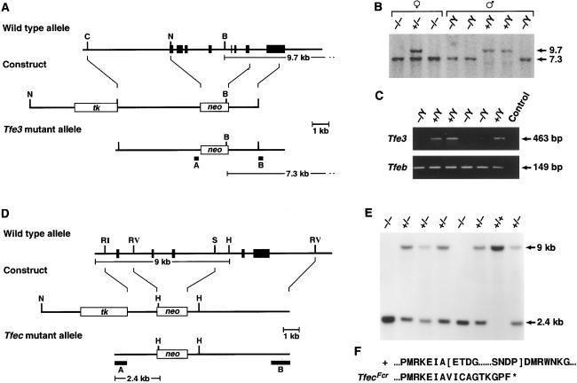
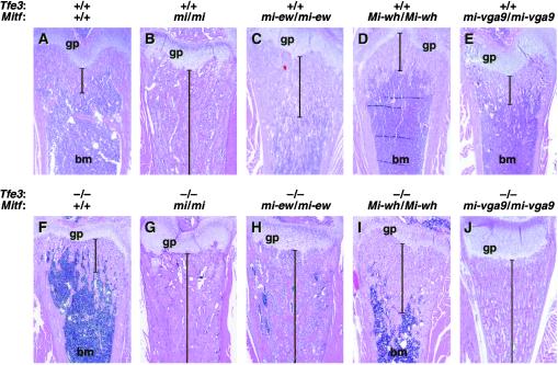
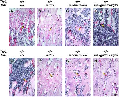
The Mitf-Tfe family of basic helix-loop-helix-leucine zipper (bHLH-Zip) transcription factors encodes four family members: Mitf, Tfe3, Tfeb, and Tfec. In vitro, each protein in the family can bind DNA as a homo- or heterodimer with other family members. Mutational studies in mice have shown that Mitf is essential for melanocyte and eye development, whereas Tfeb is required for placental vascularization. Here, we uncover a role for Tfe3 in osteoclast development, a role that is functionally redundant with Mitf. Although osteoclasts seem normal in Mitf or Tfe3 null mice, the combined loss of the two genes results in severe osteopetrosis. We also show that Tfec mutant mice are phenotypically normal, and that the Tfec mutation does not alter the phenotype of Mitf, Tfeb, or Tfe3 mutant mice. Surprisingly, our studies failed to identify any phenotypic overlap between the different Mitf-Tfe mutations. These results suggest that heterodimeric interactions are not essential for Mitf-Tfe function in contrast to other bHLH-Zip families like Myc/Max/Mad, where heterodimeric interactions seem to be essential.


Figures

Figure 1
Figure 1
Generation of Tfe3Fcr and TfecFcr mutant mice. (A) The Tfe3 knockout. The exon-intron organization of Tfe3 is shown at the top with exons indicated as black boxes. A 5.5-kb genomic fragment containing seven Tfe3 exons, the 5′ end of Tfe3, and the bHLH-Zip domains was replaced with PgkNeo. The first exon corresponds to nucleotides 14–153 of mouse Tfe3 (19). (B) Identification of Tfe3Fcr mutant animals. Tail DNA from the progeny of a heterozygous Tfe3Fcr intercross was digested with BamHI and analyzed by Southern analysis with probe B, which detects 9.7- (wild-type) and 7.3-kb (mutant) alleles. Tfe3Fcr males only transmit the mutant allele to their daughters, confirming the X chromosome linkage of Tfe3 (19). (C) Tfe3 expression in mutant animals. Kidney RNA from Tfe3Fcr and wild-type males was reversed-transcribed and PCR-amplified by using Tfe3 primers 5′-ccaagctggcttcccaggctctcac and 5′-gttaatgttgaatcgcctgcgtcg. The wild-type 463-bp fragment corresponds to nucleotides 245–708 of Tfe3 (19). The same samples were also amplified with Tfeb primers 5′-ccctgtctagcagccacctgaacg and 5′-gccgctccttggccagggctctgctc as a control. Primer pairs spanned exon-intron boundaries to eliminate false positives. (D) The Tfec knockout. A 5-kb fragment containing two Tfec bHLH exons was replaced with PgkNeo. Deleted exons correspond to nucleotides 507–639 of rat Tfec (18). (E) Identification of TfecFcr mutant animals. Tail DNA from the progeny of a heterozygous mutant intercross was digested with HindIII and analyzed by Southern analysis with probe A, which detects 9.0- (wild-type) and 2.4-kb (mutant) alleles. (F) Sequence of the TfecFcr mutant transcript. Kidney RNA from wild-type and mutant animals was reverse-transcribed and PCR-amplified with Tfec primers 5′-cagtgatgctggctgtgc and 5′-gtagccacttgatgtagtcc. Sequencing of the mutant transcript showed that it lacked two conserved Tfec functional domains (indicated by brackets in the wild-type sequence) and is out of frame for the rest of the coding region. B, BamHI; C, ClaI; N, H, HindIII; RI, EcoRI; N, NotI; RV, EcoRV; S, SacI.
Figure 2
Figure 2
Osteopetrosis in Mitf and Tfe3Fcr mutant animals. (AJ) Sections through femurs of wild-type and mutant mice showing different levels of osteopetrosis. The genotypes of the different animals are shown at the top of each image. Brackets indicate the extent of osteopetrosis. gp, growth plate; bm, bone marrow.
Figure 3
Figure 3
Osteoclast defects in Mitf and Tfe3Fcr mutant animals. (AH) Sections through femurs of wild-type and mutant mice showing different levels of osteoclast activity as determined by tartrate-resistant alkaline phosphatase staining. Arrows point to osteoclasts.

References

    1. Schreiber-Agus N, DePinho R A. BioEssays. 1998;20:808–818. - PubMed
    1. Knoepfler P S, Eisenman R N. Cell. 1999;99:447–450. - PubMed
    1. O'Hagan R C, Schreiber-Agus N, Chen K, David G, Engelman J A, Schwab R, Alland L, Thomson C, Ronning D R, Sacchettini J C, et al. Nat Genet. 2000;24:113–119. - PubMed
    1. Moore K J. Trends Genet. 1995;11:442–448. - PubMed
    1. Steingrímsson E, Tessarollo L, Reid S W, Jenkins N A, Copeland N G. Development (Cambridge, UK) 1998;125:4607–4616. - PubMed

Publication types