

Osteopontin deficiency protects joints against destruction in anti-type II collagen antibody-induced arthritis in mice
- PMID: 11930008
- PMCID: PMC123686
- DOI: 10.1073/pnas.052523599
Osteopontin deficiency protects joints against destruction in anti-type II collagen antibody-induced arthritis in mice
Abstract
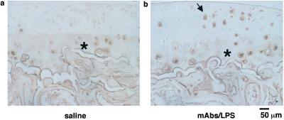
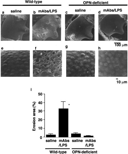
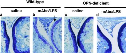
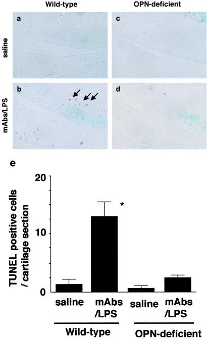
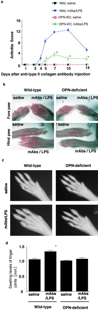
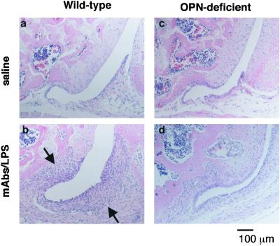
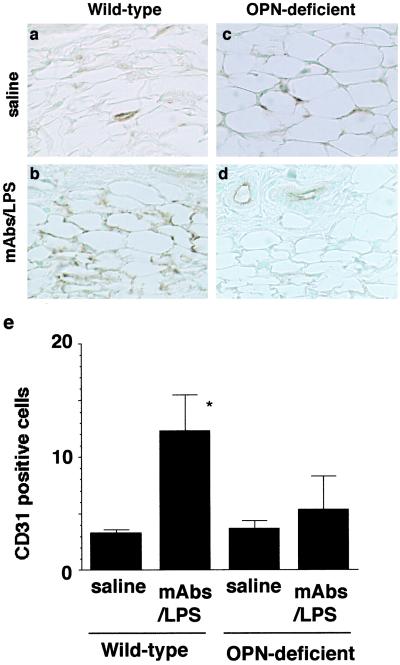
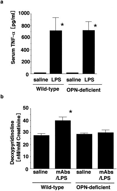
Rheumatoid arthritis is one of the most critical diseases that impair the quality of life of patients, but its pathogenesis has not yet been fully understood. Osteopontin (OPN) is an extracellular matrix protein containing Arg-Gly-Asp (RGD) sequence, which interacts with alpha(v)beta3 integrins, promotes cell attachment, and cell migration and is expressed in both synovial cells and chondrocytes in rheumatoid arthritis; however, its functional relationship to arthritis has not been known. Therefore, we investigated the roles of OPN in the pathogenesis of inflammatory process in a rheumatoid arthritis model induced by a mixture of anti-type II collagen mAbs and lipopolysaccharide (mAbs/LPS). mAbs/LPS injection induced OPN expression in synovia as well as cartilage, and this expression was associated with joint swelling, destruction of the surface structures of the joint based on scanning electron microscopy, and loss of toluidine blue-positive proteoglycan content in the articular cartilage in wild-type mice. In contrast, OPN deficiency prevented the mice from such surface destruction, loss of proteoglycan in the articular joint cartilage, and swelling of the joints even when the mice were subjected to mAbs/LPS injection. Furthermore, mAbs/LPS injection in wild-type mice enhanced the levels of CD31-positive vessels in synovia and terminal deoxynucleotidyltransferase-mediated UTP end labeling-positive chondrocytes in the articular cartilage, whereas such angiogenesis as well as chondrocyte apoptosis was suppressed significantly in OPN-deficient mice. These results indicated that OPN plays a critical role in the destruction of joint cartilage in the rheumatoid arthritis model in mice via promotion of angiogenesis and induction of chondrocyte apoptosis.
Figures
Similar articles
-
Expression of osteopontin messenger RNA and protein in rheumatoid arthritis: effects of osteopontin on the release of collagenase 1 from articular chondrocytes and synovial fibroblasts.Arthritis Rheum. 2000 Jul;43(7):1597-605. doi: 10.1002/1529-0131(200007)43:7<1597::AID-ANR25>3.0.CO;2-0. Arthritis Rheum. 2000. PMID: 10902765
-
Osteopontin Deficiency Suppresses Tumor Necrosis Factor-α-Induced Apoptosis in Chondrocytes.Cartilage. 2012 Jan;3(1):79-85. doi: 10.1177/1947603511421502. Cartilage. 2012. PMID: 26069621 Free PMC article.
-
Lack of requirement of osteopontin for inflammation, bone erosion, and cartilage damage in the K/BxN model of autoantibody-mediated arthritis.Arthritis Rheum. 2004 Aug;50(8):2685-94. doi: 10.1002/art.20381. Arthritis Rheum. 2004. PMID: 15334485
-
[Osteopontin and arthritis].Nihon Rinsho. 2003 May;61(5):887-90. Nihon Rinsho. 2003. PMID: 12755020 Review. Japanese.
-
Role of osteopontin in rheumatoid arthritis.Rheumatol Int. 2015 Apr;35(4):589-95. doi: 10.1007/s00296-014-3122-z. Epub 2014 Aug 28. Rheumatol Int. 2015. PMID: 25163663 Review.
Cited by
-
Osteopontin: A Bone-Derived Protein Involved in Rheumatoid Arthritis and Osteoarthritis Immunopathology.Biomolecules. 2023 Mar 9;13(3):502. doi: 10.3390/biom13030502. Biomolecules. 2023. PMID: 36979437 Free PMC article. Review.
-
Osteopontin expression in the brain triggers localized inflammation and cell death when immune cells are activated by pertussis toxin.Mediators Inflamm. 2014;2014:358218. doi: 10.1155/2014/358218. Epub 2014 Nov 24. Mediators Inflamm. 2014. PMID: 25525298 Free PMC article.
-
Molecular targets in arthritis and recent trends in nanotherapy.Int J Nanomedicine. 2015 Aug 26;10:5407-20. doi: 10.2147/IJN.S89156. eCollection 2015. Int J Nanomedicine. 2015. PMID: 26345140 Free PMC article. Review.
-
Regulation of T-helper-cell lineage development by osteopontin: the inside story.Nat Rev Immunol. 2009 Feb;9(2):137-41. doi: 10.1038/nri2460. Nat Rev Immunol. 2009. PMID: 19096390 Free PMC article. Review.
-
Danger signal extracellular calcium initiates differentiation of monocytes into SPP1/osteopontin-producing macrophages.Cell Death Dis. 2022 Jan 12;13(1):53. doi: 10.1038/s41419-022-04507-3. Cell Death Dis. 2022. PMID: 35022393 Free PMC article.
References
Publication types
MeSH terms
Substances
Grants and funding
LinkOut - more resources
Full Text Sources
Other Literature Sources
Medical
Research Materials
