Review
. 2002 Apr;15(2):194-222.
doi: 10.1128/CMR.15.2.194-222.2002.

Lung infections associated with cystic fibrosis

Affiliations
Free PMC article
Review

Lung infections associated with cystic fibrosis

Jeffrey B Lyczak et al. Clin Microbiol Rev. 2002 Apr.
Free PMC article

Abstract

While originally characterized as a collection of related syndromes, cystic fibrosis (CF) is now recognized as a single disease whose diverse symptoms stem from the wide tissue distribution of the gene product that is defective in CF, the ion channel and regulator, cystic fibrosis transmembrane conductance regulator (CFTR). Defective CFTR protein impacts the function of the pancreas and alters the consistency of mucosal secretions. The latter of these effects probably plays an important role in the defective resistance of CF patients to many pathogens. As the modalities of CF research have changed over the decades from empirical histological studies to include biophysical measurements of CFTR function, the clinical management of this disease has similarly evolved to effectively address the ever-changing spectrum of CF-related infectious diseases. These factors have led to the successful management of many CF-related infections with the notable exception of chronic lung infection with the gram-negative bacterium Pseudomonas aeruginosa. The virulence of P. aeruginosa stems from multiple bacterial attributes, including antibiotic resistance, the ability to utilize quorum-sensing signals to form biofilms, the destructive potential of a multitude of its microbial toxins, and the ability to acquire a mucoid phenotype, which renders this microbe resistant to both the innate and acquired immunologic defenses of the host.


Figures

FIG. 1.
FIG. 1.
Diagram of a sweat gland, showing paths taken by chloride ions (arrows) during secretion. In both normal and CF sweat glands in the dermis, chloride is present in secretions at a concentration of 105 mEq, equaling that in serum (“isotonic”). (Top) In the normal sweat gland, chloride is absorbed out of the sweat in a CFTR-dependent manner as the sweat travels from the gland to the skin surface. As a result, the chloride concentration in normal sweat is below that in serum (“hypotonic”), with <40 mEq considered normal and <20 mEq being typical. (Bottom) In the CF sweat gland, chloride absorption is hindered by defective CFTR function. As a result, sweat which reaches the skin surface has higher than normal chloride concentrations (>60 mEq).
FIG. 2.
FIG. 2.
Chloride secretion in a pulmonary secretory epithelial cell. The ability of secretory epithelia to secrete fluid rests on the energy provided by the ubiquitous, basolaterally located Na+/K+-ATPase (Step 1), which maintains a low intracellular concentration of Na+ by actively pumping it out of the cell. The low intracellular Na+ concentration, coupled with the high extracellular Na+ concentration and the negative transmembrane potential, drives the passive diffusion of Na+ into the cell down the concentration gradient. The channel through which this passive diffusion occurs (the Na+/K+/2Cl cotransporter) requires concomitant transport of Na+, K+, and Cl for any transport to occur at all. Thus, the passive diffusion of Na+ into the cell is coupled with an intracellular accumulation of Cl against its electrochemical gradient (Step 2). When the cell is stimulated to secrete, Cl channels open, allowing Cl to exit down its electrochemical gradient (Step 3). Sodium ions follow the Cl ions through a paracellular pathway, and water follows the salt due to the resulting osmotic gradient (Step 4).
FIG. 3.
FIG. 3.
CFTR regulates apical ion transport via several mechanisms. The first of these mechanisms is the innate function of CFTR as a chloride channel (triangle 1). Second, the R domain of CFTR associates with and regulates the activity of the potassium channel ROMK2 (triangle 2). Third, CFTR mediates transport of ATP across the plasma membrane. This extracellular ATP can then bind to the purinergic receptor (PY2R), which regulates the activity of the ORCC (triangle 3). Lastly, there is evidence that CFTR can directly activate the chloride import activity of the ORCC and repress the sodium channel ENaC (triangle 4). The plasma membrane shown in the figure represents the surface of a generic epithelial cell, with characteristics of epithelia from several tissues.
FIG. 4.
FIG. 4.
Schematic diagram of the proposed structure of CFTR. A member of the ABC family, CFTR consists of a tandem repeat of the ABC motif. This motif comprises a membrane-spanning domain (composed of six transmembrane stretches of amino acids) followed by an NBD. In CFTR, the two occurrences of this motif are separated by a regulatory (R) domain. Each NBD is able to bind and hydrolyze ATP to operate chloride channel function: hydrolysis of ATP by NBD-1 opens the chloride channel, while ATP hydrolysis by NBD-2 closes the channel. Channel function is further regulated by phosphorylation of serine residues in the R domain.
FIG. 5.
FIG. 5.
Correlation in 11 European countries of prevalence of the ΔF508 CFTR allele with the incidence of S. enterica serovar Typhi infection 23 years earlier.
FIG. 6.
FIG. 6.
(A) Comparison of mucociliary clearance in the normal airway and the CF airway. The normal airway epithelium is covered with a biphasic mucus layer consisting of a viscous upper layer and a more fluid lower (periciliary) layer. Concerted beating of epithelial cell cilia causes the mucus to flow unidirectionally toward the esophagus, carrying with it any microorganisms which become trapped in the mucus. In the CF airway, alterations in either mucus secretion, mucus reabsorption, or both cause the mucus layer to become uniformly viscous, such that beating of the epithelial cilia is no longer sufficient to propel the mucus toward the esophagus. Bacteria can therefore persist in the airway. Defects in the CFTR protein can also increase the adhesion of P. aeruginosa to the airway epithelium. (B) CFTR-dependent internalization of P. aeruginosa causes apoptosis and desquamation of bacteria-laden epithelial cells. In the normal airway, these apoptotic cells and the bacteria they contain are probably removed from the airway via the mucociliary escalator. In the CF airway, this clearance mechanism does not function normally due to inefficient internalization of bacteria stemming from a lack of CFTR protein. It is possible that apoptotic bodies derived from desquamated epithelial cells are later phagocytosed by dendritic cells for subsequent processing and presentation of bacterial antigens to T lymphocytes.
FIG. 6.
FIG. 6.
(A) Comparison of mucociliary clearance in the normal airway and the CF airway. The normal airway epithelium is covered with a biphasic mucus layer consisting of a viscous upper layer and a more fluid lower (periciliary) layer. Concerted beating of epithelial cell cilia causes the mucus to flow unidirectionally toward the esophagus, carrying with it any microorganisms which become trapped in the mucus. In the CF airway, alterations in either mucus secretion, mucus reabsorption, or both cause the mucus layer to become uniformly viscous, such that beating of the epithelial cilia is no longer sufficient to propel the mucus toward the esophagus. Bacteria can therefore persist in the airway. Defects in the CFTR protein can also increase the adhesion of P. aeruginosa to the airway epithelium. (B) CFTR-dependent internalization of P. aeruginosa causes apoptosis and desquamation of bacteria-laden epithelial cells. In the normal airway, these apoptotic cells and the bacteria they contain are probably removed from the airway via the mucociliary escalator. In the CF airway, this clearance mechanism does not function normally due to inefficient internalization of bacteria stemming from a lack of CFTR protein. It is possible that apoptotic bodies derived from desquamated epithelial cells are later phagocytosed by dendritic cells for subsequent processing and presentation of bacterial antigens to T lymphocytes.
FIG. 7.
FIG. 7.
The AlgU alternative sigma factor (also called AlgT) directly regulates expression of itself, of the alginate biosynthetic operon, and of several other regulatory genes. The figure depicts events leading to transcriptional regulation of the algD gene (encoding GDP-mannose dehydrogenase), which lies at the 5′ end of the alginate biosynthetic operon. The activity of the AlgU transcription factor is negatively regulated by the two anti-sigma factors MucA and MucB (Tier 1). When AlgU remains in an active state, it is then able to initiate transcription of the algB and algR genes, which encode transcriptional regulatory proteins (Tier 2). These transcriptional activators then positively regulate algD transcription. Regulation of algD by AlgB and AlgR may be further modified by other events such as phosphorylation of AlgB and AlgR and dimerization of AlgR with another transactivator, AlgZ (Tier 3). Symbols: formula image
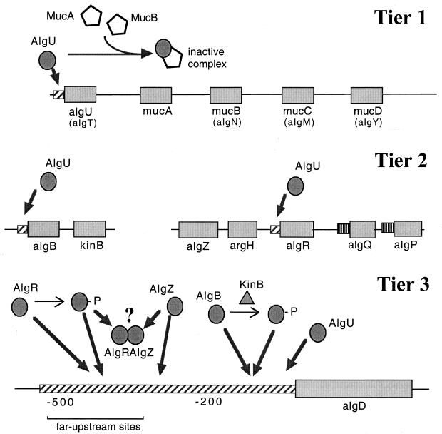
, constitutive (σ70-like) promoter; formula image, heat shock (σ54-like) promoter; formula image, transcriptional activator; formula image, transcriptional inhibitor; formula image, structural gene; ➞, positive regulation; →, posttranslational modification; formula image, kinase; -P, phosphate.

References

    1. Reference deleted.
    1. Albert, M. L., S. F. Pearce, L. M. Francisco, B. Sauter, P. Roy, R. L. Silverstein, and N. Bhardwaj. 1998. Immature dendritic cells phagocytose apoptotic cells via alphavbeta5 and CD36, and cross-present antigens to cytotoxic T lymphocytes. J. Exp. Med. 188:1359-1368. - PMC - PubMed
    1. Albus, A. M., E. C. Pesci, L. J. Runyen-Janecky, S. E. West, and B. H. Iglewski. 1997. Vfr controls quorum sensing in Pseudomonas aeruginosa. J. Bacteriol. 179:3928-3935. - PMC - PubMed
    1. Allan, J. D., A. Mason, and A. D. Moss. 1973. Nutritional supplementation in treatment of cystic fibrosis of the pancreas. Am. J. Dis. Child. 126:22-26. - PubMed
    1. Allewelt, M., F. T. Coleman, M. Grout, G. P. Priebe, and G. B. Pier. 2000. Acquisition of expression of the Pseudomonas aeruginosa ExoU cytotoxin leads to increased bacterial virulence in a murine model of acute pneumonia and systemic spread. Infect. Immun. 68:3998-4004. - PMC - PubMed

Publication types

MeSH terms

Substances