Review
. 2002 Apr;15(2):310-26.
doi: 10.1128/CMR.15.2.310-326.2002.

Angiogenic effects of extracellular human immunodeficiency virus type 1 Tat protein and its role in the pathogenesis of AIDS-associated Kaposi's sarcoma

Affiliations
Review

Angiogenic effects of extracellular human immunodeficiency virus type 1 Tat protein and its role in the pathogenesis of AIDS-associated Kaposi's sarcoma

Giovanni Barillari et al. Clin Microbiol Rev. 2002 Apr.

Abstract

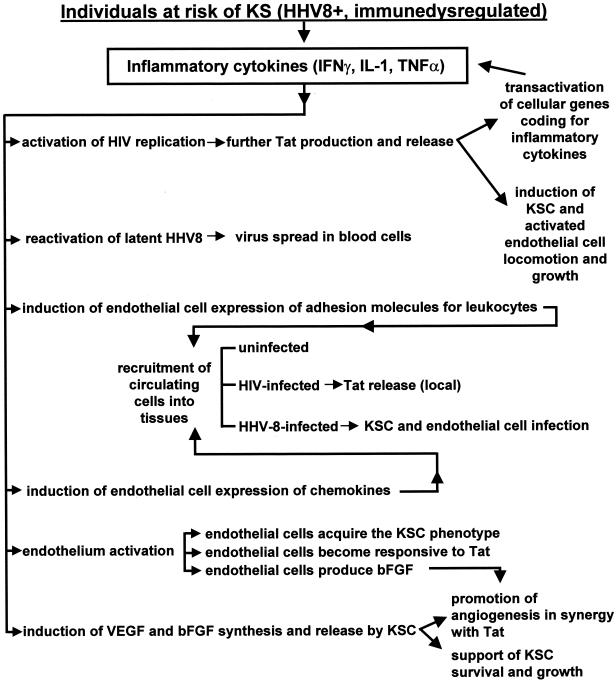
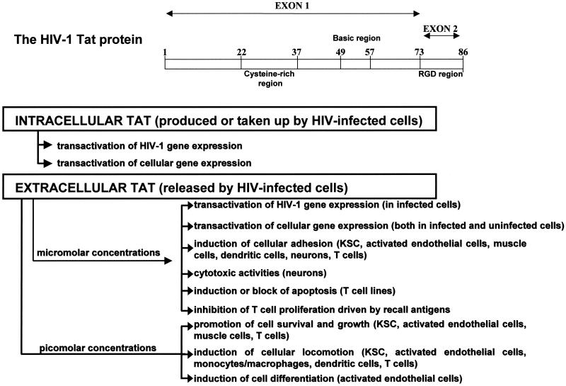
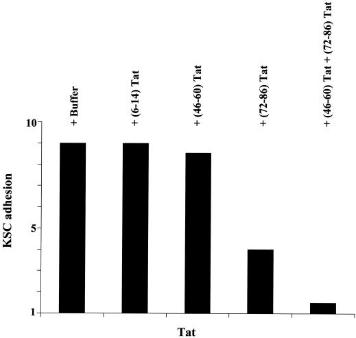
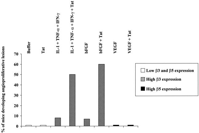
The Tat protein of human immunodeficiency virus (HIV) type 1 is a transactivator of viral gene expression that is required for virus replication and spread. Moreover, Tat is released by acutely HIV-infected cells via a leaderless secretory pathway and in a biologically active form that exerts effects on both HIV-infected and uninfected cells from different organs and systems. This review focuses on the activities of extracellular Tat protein on endothelial cells, on angiogenesis, and on the pathogenesis of AIDS-associated angioproliferative diseases such as Kaposi's sarcoma. In particular, we discuss results from different groups indicating that Tat mimics the proangiogenic activities of extracellular matrix molecules and that it enhances the effects of angiogenic factors.


Figures

FIG. 1.
FIG. 1.
Interplay among HHV-8, inflammatory cytokines, angiogenic factors and HIV-1 Tat protein in AIDS-KS pathogenesis.
FIG. 2.
FIG. 2.
Structure and biological properties of HIV-1 Tat protein.
FIG. 3.
FIG. 3.
KSC adhesion to Tat is mediated by the RGD region of the protein, whereas Tat basic sequence facilitates this interaction. KSC were suspended by trypsinization and preincubated for 90 min at 4°C with rotation and with one of the following competitor peptides (at 10 μg/ml): (6-14)Tat, a peptide spanning the Tat N-terminal region; (46-60)Tat, a peptide spanning the Tat basic region; or (72-86)Tat, a peptide spanning the Tat C-terminal region and containing the RGD sequence (all from American Biotechnologies Inc.). After incubation, KSC were seeded on plastic plates (Flow Laboratories) coated with recombinant Tat protein (30 μg/cm2) and incubated for 1 h at 37°C in a CO2 atmosphere. Nonadherent cells were removed by washing the plates with phosphate-buffered saline solution, whereas adherent cells were fixed, stained, and quantitated as previously described (16, 28). The number of adherent cells is expressed as the fold increase in KSC attachment compared to the adhesion seen with of bovine serum albumin (30 μg/cm2). This did not induce KSC attachment and was given a unitary value.
FIG. 4.
FIG. 4.
Combined IL-1β, TNF-α, and IFN-γ or bFGF, but not VEGF, synergizes with Tat in inducing the development of angioproliferative, KS-like lesions in nude mice. Nude mice received subcutaneous injections with recombinant IL-1β, TNF-α, and IFN-γ (0.5 μg of each), bFGF (0.1 μg), VEGF (1 μg), or Tat (10 μg), alone or combined. Results are expressed as the percentage of mice developing macroscopic angioproliferative lesions 6 days after protein inoculation (18, 54). Eight to twelve mice were inoculated per experimental condition. Results from staining experiments performed with rabbit polyclonal antibodies by the peroxidase-antiperoxidase method (18, 54) on frozen tissues from nude mice injected with the above-mentioned cytokines are expressed as β3 or β5 expression level increases over the expression of these integrins in tissues from mice injected with protein resuspension buffer (phosphate-buffered saline solution-0.1% bovine serum albumin) (18).

References

    1. Aboulafia, D., S. A. Miles, S. R. Saks, and R. T. Mitsuyasu. 1989. Intravenous recombinant tumor necrosis factor in the treatment of AIDS-related Kaposi's sarcoma. J. Acquir. Immune Defic. Syndr. 2:54-58. - PubMed
    1. Albini, A., G. Barillari, R. Benelli, R. C. Gallo, and B. Ensoli. 1995. Angiogenic properties of human immunodeficiency virus type 1 Tat protein. Proc. Natl. Acad. Sci. USA 92:4838-4842. - PMC - PubMed
    1. Albini, A., R. Soldi, D. Giunciuglio, E. Giraudo, R. Benelli, L. Primo, D. Noonan, M. Salio, G. Camussi, W. Rockl, and F. Bussolino. 1996. The angiogenesis induced by HIV-1 tat protein is mediated by the Flk-1/KDR receptor on vascular endothelial cells. Nat. Med. 2:1371-1375. - PubMed
    1. Albini, A., R. Benelli, M. Presta, M. Rusnati, M. Ziche, A. Rubartelli, G. Paglialunga, F. Bussolino, and D. Noonan. 1996. HIV-1 tat protein is a heparin binding angiogenic growth factor. Oncogene 12:289-297. - PubMed
    1. Albini, A., R. Benelli, D. Giunciuglio, T. Cai, G. Mariani, S. Ferrini, and D. Noonan. 1998. Identification of a novel domain of HIV tat involved in monocyte chemotaxis. J. Biol. Chem. 273:15895-15900. - PubMed

Publication types

MeSH terms

Substances