. 2002 May;76(9):4275-86.
doi: 10.1128/jvi.76.9.4275-4286.2002.

Avian bic, a gene isolated from a common retroviral site in avian leukosis virus-induced lymphomas that encodes a noncoding RNA, cooperates with c-myc in lymphomagenesis and erythroleukemogenesis

Affiliations

Avian bic, a gene isolated from a common retroviral site in avian leukosis virus-induced lymphomas that encodes a noncoding RNA, cooperates with c-myc in lymphomagenesis and erythroleukemogenesis

Wayne Tam et al. J Virol. 2002 May.

Abstract

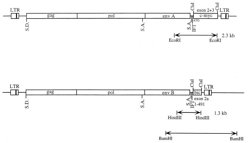
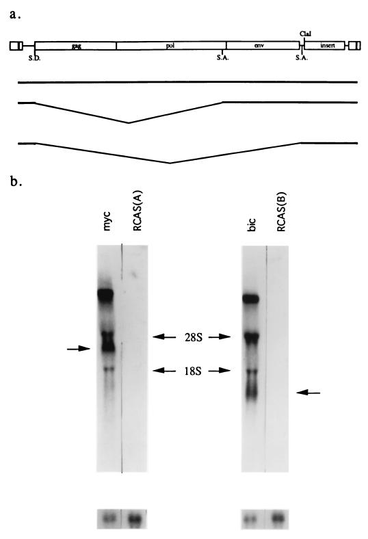
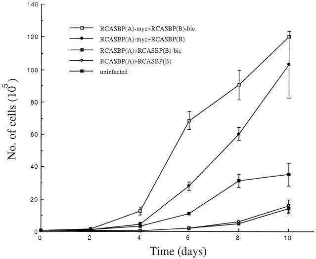
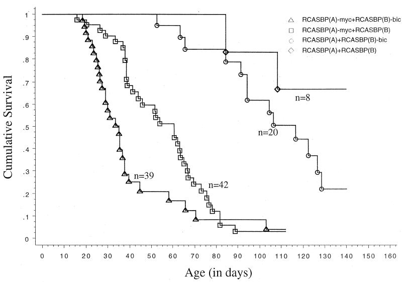
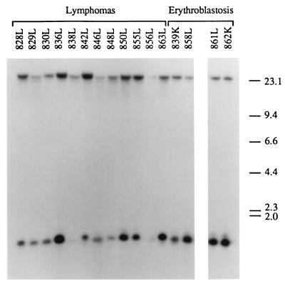
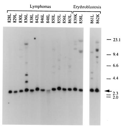
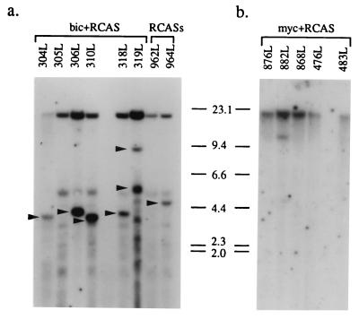
bic is a novel gene identified at a common retroviral integration site in avian leukosis virus-induced lymphomas and has been implicated as a collaborator with c-myc in B lymphomagenesis. It lacks an extensive open reading frame and is believed to function as an untranslated RNA (W. Tam, Gene 274:157-167, 2001; W. Tam, D. Ben-Yehuda, and W. S. Hayward, Mol. Cell. Biol. 17:1490-1502, 1997). The oncogenic potential of bic, particularly its ability to cooperate with c-myc in oncogenesis, was tested directly by expressing c-myc and bic, either singly or in pairwise combination, in cultured chicken embryo fibroblasts (CEFs) and in chickens using replication-competent retrovirus vectors. Coexpression of c-myc and bic in CEFs caused growth enhancement of cells. Most importantly, chick oncogenicity assays demonstrated that bic can cooperate with c-myc in lymphomagenesis and erythroleukemogenesis. The present study provides direct evidence for the involvement of untranslated RNAs in oncogenesis and provides further support for the role of noncoding RNAs as riboregulators.


Figures

FIG. 1.
FIG. 1.
Structures of the RCASBP(A)-myc and RCASBP(B)-bic proviruses. c-myc and bic cDNA sequences were inserted into the ClaI sites of the replication-competent retrovirus vectors RCASBP(A) and RCASBP(B), respectively. The c-myc insert consists of chicken c-myc exons 2 and 3. The bic insert comprises nucleotides 1 to 491 of bic exon 2. The virus internal myc-specific (2.3-kb EcoRI) and bic-specific (1.3-kb HindIII) fragments are shown. The BamHI RCASBP(B)-bic virus-cell junction fragment is also depicted. S.D., splice donor; S.A., splice acceptor; IFT, in-frame terminator.
FIG. 2.
FIG. 2.
Expression of c-myc and bic in CEFs using RCASBP(A)-myc and RCASBP(B)-bic. (a) Schematic of expected RNAs generated from RCASBP(A)-myc and RCASBP(B)-bic constructs. These vectors express cDNA inserts as spliced subgenomic mRNA. SD, splice donor site; SA, splice acceptor site. (b) Northern blot analysis of c-myc and bic expression. Total RNA derived from CEFs mass-infected with RCASBP(A)-myc (lane myc), RCASBP(A) [lane RCAS(A)], RCASBP(B)-bic (lane bic), or RCASBP(B) [lane RCAS(B)] were probed with either a radiolabeled c-myc exon 3 cDNA probe (left) or a bic exon 2 probe (right). The subgenomic mRNA expressing c-myc or bic is indicated by an arrow. The blot was stripped and rehybridized to a chicken actin probe as a control for loading. The radioactive bands observed at the 28S and 18S regions are probably due to hybridization of probe sequence to viral RNAs that comigrated nonspecifically with the rRNAs.
FIG. 3.
FIG. 3.
Expression of c-myc and bic in doubly infected CEFs. (a) CEF cultures were simultaneously infected with two viruses as indicated, and total RNA from these cultures was subjected to Northern analysis as described in Materials and Methods. myc plus bic, RCASBP(A)-myc and RCASBP(B)-bic; myc plus RCAS, RCASBP(A)-myc and RCASBP(B); bic plus RCAS, RCASBP(A) and RCASBP(B)-bic; RCASs, RCASBP(A) and RCASBP(B). (b) Protein lysates prepared from doubly infected CEF cultures were separated by electrophoresis and subjected to Western blot analysis using monoclonal antibodies against c-Myc. The p55 myc protein expressed by RCASBP(A)-myc is indicated. The lanes are designated as in a. The faster-migrating bands represent cross-reactive proteins.
FIG. 4.
FIG. 4.
Enhanced growth of CEFs overexpressing c-myc and bic. CEFs doubly infected with the indicated viruses were plated at a starting density of 105 cells/plate. The cultures were trypsinized and counted at the indicated time points. The experiment was done in duplicate. The error bars represent the standard deviation at each time point.
FIG. 5.
FIG. 5.
Kaplan-Meier survival curves (neoplastic diseases only) for animals infected with different retroviruses. Eighteen-day chick embryos were doubly infected with retrovirus vectors as indicated. Animals inoculated with different viruses were maintained in separate cages and observed for disease. Each death event (indicated by a vertical line) represents the death of an animal due to neoplastic disease.
FIG. 6.
FIG. 6.
Detection of proviral integrations of RCASBP(B)-bic in lymphomas and erythroblastosis tumors. High-molecular-weight DNA isolated from the tumor tissues (L, liver; K, kidney) of birds infected with RCASBP(A)-myc and RCASBP(B)-bic were digested with HindIII and subjected to Southern blot analysis using a radiolabeled bic exon 2a probe (see Fig. 1). The band of lower mobility represents the endogenous bic gene restriction fragment. Fragment sizes are indicated in kilobases.
FIG. 7.
FIG. 7.
Detection of RCASBP(B)-bic virus junction fragments in lymphomas and erythroblastosis tumors. BamHI-digested tumor DNA was hybridized to a radiolabeled bic exon 2a probe in Southern blot analysis (see Fig. 1). The 2.5-kb endogenous BamHI fragment is indicated by an arrow. Bands of lower mobilities represent virus junction fragments. Sizes are shown in kilobases.
FIG. 8.
FIG. 8.
Detection of c-myc and bic gene arrangements in lymphomas. (a) Lymphoma DNA from animals doubly infected with RCASBP(A) and RCASBP(B)-bic (bic plus RCAS) or RCASBP(A) plus RCASBP(B) (RCASs) was digested with EcoRI and hybridized to a radiolabeled c-myc exon 3 probe in Southern blot analysis. The 15-kb germ line EcoRI fragment is present in all samples. Bands which represent rearranged c-myc alleles are indicated by arrowheads. The multiple bands of similar sizes which are observed in all the lanes are likely due to nonspecific hybridization of probe sequences. (b) High-grade lymphoma DNA from animals infected with RCASBP(A)-myc and RCASBP(B) (myc plus RCAS) was digested with HindIII, and Southern hybridization was performed using a radiolabeled bic exon 2a probe. The 23-kb endogenous HindIII fragment is present in all samples. The additional fragment observed in lane 882L represents a rearranged bic allele.

References

    1. Adams, J. M., and S. Cory. 1992. Oncogene cooperation in leukaemogenesis. Cancer Surv. 15:119-141. - PubMed
    1. Adams, J. M., A. W. Harris, A. Strasser, S. Ogilvy, and S. Cory. 1999. Transgenic models of lymphoid neoplasia and development of a pan-hematopoietic vector. Oncogene 18:5268-5277. - PubMed
    1. Alexander, W. S., O. Bernard, S. Cory, and J. M. Adams. 1989. Lymphomagenesis in E mu-myc transgenic mice can involve ras mutations. Oncogene 4:575-581. - PubMed
    1. Askew, D. S., and F. Xu. 1999. New insights into the function of noncoding RNA and its potential role in disease pathogenesis. Histol. Histopathol. 14:235-241. - PubMed
    1. Baba, T. W., and E. H. Humphries. 1985. Formation of a transformed follicle is necessary but not sufficient for development of an avian leukosis virus-induced lymphoma. Proc. Natl. Acad. Sci. USA 82:213-216. - PMC - PubMed

Publication types

MeSH terms