. 2002 May;76(9):4312-20.
doi: 10.1128/jvi.76.9.4312-4320.2002.

Involvement of the matrix protein in attachment of porcine reproductive and respiratory syndrome virus to a heparinlike receptor on porcine alveolar macrophages

Affiliations

Involvement of the matrix protein in attachment of porcine reproductive and respiratory syndrome virus to a heparinlike receptor on porcine alveolar macrophages

P L Delputte et al. J Virol. 2002 May.

Abstract

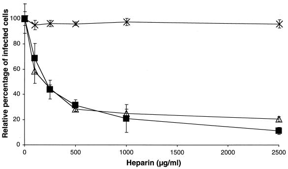
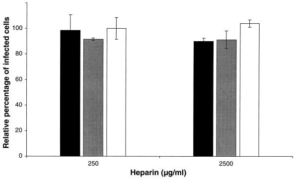
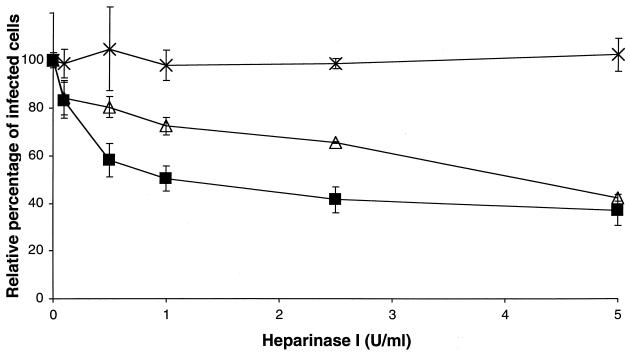
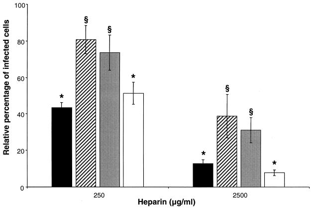
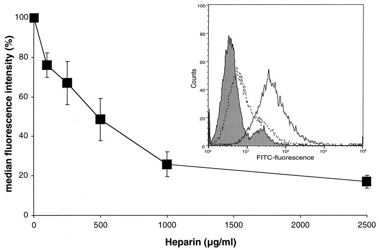
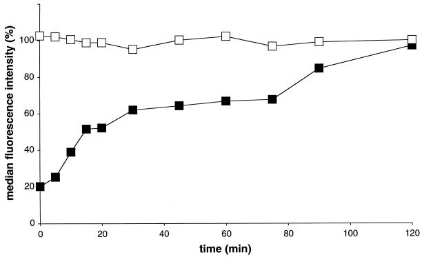
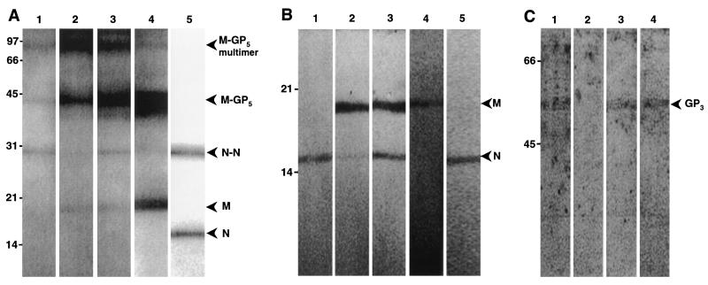
The porcine reproductive and respiratory syndrome virus (PRRSV) has a very restricted tropism for well-differentiated cells of the monocyte-macrophage lineage, which is probably determined by specific receptors on these cells. In this study, the importance of heparinlike molecules on porcine alveolar macrophages (PAM) for PRRSV infection was determined. Heparin interacted with the virus and reduced infection of PAM up to 92 or 88% for the American and European types of PRRSV, respectively. Other glycosaminoglycans, similar to heparin, had no significant effect on infection while heparinase treatment of PAM resulted in a significant reduction of the infection. Analysis of infection kinetics showed that PRRSV attachment to heparan sulfate occurs early in infection. A heparin-sensitive binding step was observed which converted completely into a heparin-resistant binding after 120 min at 4 degrees C. Using heparin-affinity chromatography and sodium dodecyl sulfate-polyacrylamide gel electrophoresis (SDS-PAGE), it was observed that the structural matrix (M) and nucleocapsid (N) proteins attached to heparin. Nonreducing SDS-PAGE revealed that M bound to heparin mainly as a complex with glycoprotein GP(5) and that the N protein bound to heparin as a homodimer. GP(3), which was identified as a minor structural protein of European types of PRRSV, did not bind to heparin. Since the N protein is not exposed on the virion surface, it was concluded that the structural M protein and the M-GP(5) complex contribute to PRRSV attachment on a heparinlike receptor on PAM. This is the first report that identifies a PRRSV ligand for a cell surface heparinlike receptor on PAM.


Figures

FIG. 1.
FIG. 1.
Effect of heparin on PRRSV infection of PAM. PRRSV (▪), PrV (▵), and PrV gCnull (×) were incubated for 1 h at 37°C with different concentrations of heparin. PAM were inoculated with the mixtures, washed after 2 h of incubation at 37°C to remove unbound virus, and finally fixed after 10 h. The data represent the means ± standard deviations (error bars) of triplicate assays.
FIG. 2.
FIG. 2.
Effect of incubation of PAM with heparin before infection. PAM were incubated for 1 h at 37°C with heparin, washed extensively, and incubated with PRRSV (solid bars), PrV (shaded bars), and PrV gC (open bars). The cells were washed after 2 h of incubation to remove unbound virus, and the cells were fixed after 10 h. The data represent the means ± standard deviations (error bars) of triplicate assays. No significant differences were observed in comparison to the control (no heparin added).
FIG. 3.
FIG. 3.
Effect of heparinase treatment of PAM on PRRSV infection. Heparinase-treated PAM were washed and incubated with PRRSV (▪), PrV (▵), and PrV gCnull (×). The PAM were washed after 2 h at 37°C and fixed after a total infection period of 10 h. The data represent the means ± standard deviations (error bars) of triplicate assays.
FIG. 4.
FIG. 4.
Effect of other GAG on PRRSV infection. Virus was incubated for 1 h at 37°C with a 2,500-μg/ml concentration of chondroitin sulfate A (▪) or dermatan sulfate (□). PAM were incubated with the virus-GAG mixtures, washed after 2 h, and fixed after a total infection time of 10 h. The data represent the means ± standard deviations of triplicate assays. No significant differences were observed in comparison to the control (no GAG added).
FIG. 5.
FIG. 5.
Effect of heparin on different PRRSV strains. PRRSV 94V360 (solid bars), LV (striped bars), US-5 (shaded bars), and VR-2332 (open bars) were incubated for 1 h at 37°C with different concentrations of heparin. PAM were inoculated with the mixtures, washed after 2 h of incubation at 37°C to remove unbound virus, and finally fixed after a total infection time of 10 h. The data represent the means ± standard deviations (error bars) of triplicate assays. The analysis of variance test and the least-significant-difference post hoc test were performed to compare the sensitivities of the different PRRSV strains to heparin, with heparin at both at 250 and 2,500 μg/ml. A different symbol on top of two bars indicates that the number of infected cells of these two PRRSV strains are significantly different at the given concentration of heparin (P < 0.01).
FIG. 6.
FIG. 6.
Heparin inhibits PRRSV attachment to PAM in a dose-dependent manner. Biotinylated PRRSV was incubated with different concentrations of heparin, and PAM were inoculated with the mixtures at 4°C for 1 h. After washing, bound virus particles were stained by direct immunofluorescence, and fluorescence intensity was analyzed by flow cytometry. The data represent the means ± standard deviations (error bars) of triplicate assays. The histogram shows the green fluorescence intensity frequency when no virus was added (shaded histogram), when virus was added (solid-line histogram), and when virus and heparin was added (dotted-line histogram).
FIG. 7.
FIG. 7.
Heparin-dependent kinetics of PRRSV attachment. PAM were inoculated with PRRSV at 4°C, unbound virus was removed by washing the cells after a 5-min incubation at 4°C, and at different time intervals PAM were washed with PBS-F with (▪) or without (□) heparin (2,500 μg/ml). Attachment was evaluated at 4°C by immunofluorescent staining of bound virus particles with streptavidin-FITC and flow cytometric analysis. The data represent the means of duplicate assays.
FIG. 8.
FIG. 8.
PRRSV (▪), PrV (▵), and PrV gCnull (×) were incubated with different concentrations of heparin Sepharose for 1 h at 37°C. The mixtures were centrifuged and the supernatant was titrated. The data are shown relatively to the original TCID50 of each virus suspension and represent the mean values of duplicate experiments.
FIG. 9.
FIG. 9.
SDS-PAGE analysis of the binding of PRRSV proteins to heparin Sepharose. PRRSV proteins were solubilized with Tris buffer containing 1% NP-40. (A) Analysis of bound fractions and original lysates under nonreducing conditions. Solubilized proteins were added to a heparin Sepharose column, and bound fractions were eluted with PBS containing heparin at 4 mg/ml (PBS-H) (lane 1), with PBS-H containing 1.5 M NaCl (lane 2), or with PBS-H containing 2 M NaCl (lane 3). Proteins were detected on a WB with a mixture of MAbs directed against the nucleocapsid (N) and the matrix (M) protein. Original lysates were also analyzed with anti-M MAb (lane 4) and anti-N MAb (lane 5). (B) Lanes 1 to 5 contain the same samples as lanes 1 to 5 in panel A, except that they were analyzed under reducing conditions. (C) Detection of PRRSV GP3 in different fractions obtained. The original lysate (lane 1), the bound fraction (lane 2), the unbound fraction (lane 3), and the wash fractions (lane 4) were analyzed for the presence of GP3 under reducing conditions. Molecular weight standards (in thousands) are indicated on the left of each blot, and positions of different PRRSV proteins and protein complexes are indicated with an arrowhead.

References

    1. Benfield, D. A., E. Nelson, J. E. Collins, L. Harris, S. M. Goyal, D. Robinson, W. T. Christianson, R. B. Morrison, D. Gorcyca, and D. Chladek. 1992. Characterisation of swine infertility and respiratory syndrome (SIRS) virus (isolate ATCC VR-2332). J. Vet. Diagn. Investig. 4:127-133. - PubMed
    1. Bernfield, M., M. Götte, P. W. Park, O. Reizes, M. L. Fitzgerald, J. Lincecum, and M. Zako. 1999. Functions of cell surface heparan sulphate proteoglycans. Annu. Rev. Biochem. 68:729-777. - PubMed
    1. Byrnes, A. P., and D. E. Griffin. 1998. Binding of Sindbis virus to cells surface heparan sulfate. J. Virol. 72:7349-7356. - PMC - PubMed
    1. Cavanagh, D. 1997. Nidovirales: a new order comprising Coronaviridae and Arteriviridae. Arch. Virol. 142:629-633. - PubMed
    1. Collins, J. E., D. A. Benfield, W. T. Christianson, L. Harris, J. C. Hennings, D. P. Shaw, S. M. Goyal, S. McCullough, R. B. Morrison, H. S. Joo, D. Gorcyca, and D. Chladek. 1992. Isolation of swine infertility and respiratory syndrome virus (isolate ATCC VR-2332) in North America and experimental reproduction of the disease in gnotobiotic pigs. J. Vet. Diagn. Investig. 4:117-126. - PubMed

Publication types

MeSH terms

LinkOut - more resources