. 2002 May;76(9):4567-79.
doi: 10.1128/jvi.76.9.4567-4579.2002.

The oncogenic protein kinase Tpl-2/Cot contributes to Epstein-Barr virus-encoded latent infection membrane protein 1-induced NF-kappaB signaling downstream of TRAF2

Affiliations

The oncogenic protein kinase Tpl-2/Cot contributes to Epstein-Barr virus-encoded latent infection membrane protein 1-induced NF-kappaB signaling downstream of TRAF2

Aristides G Eliopoulos et al. J Virol. 2002 May.

Abstract

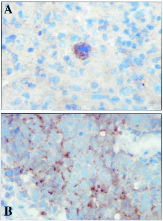
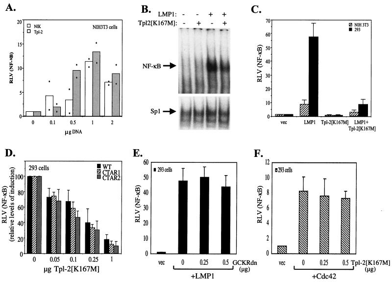
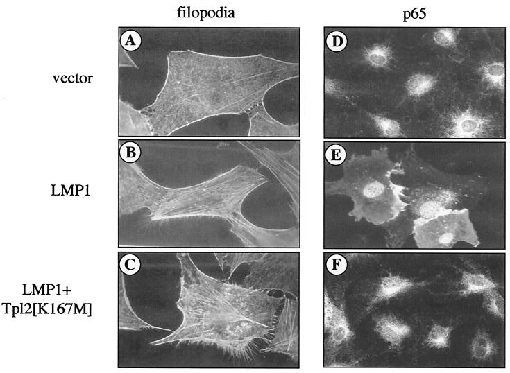
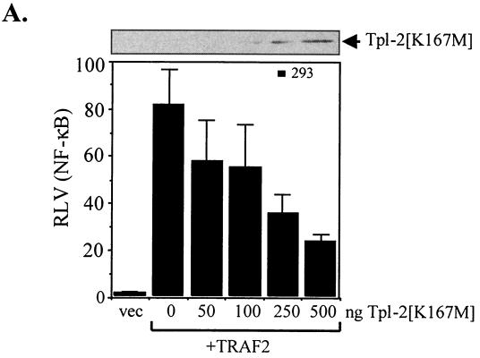
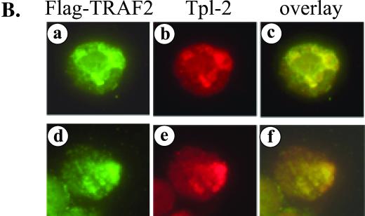
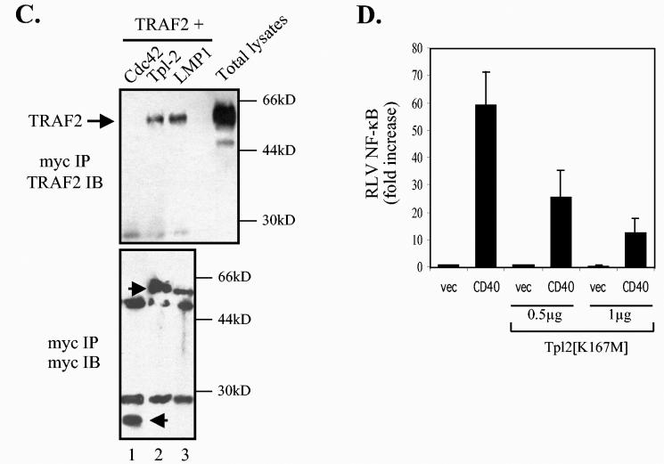
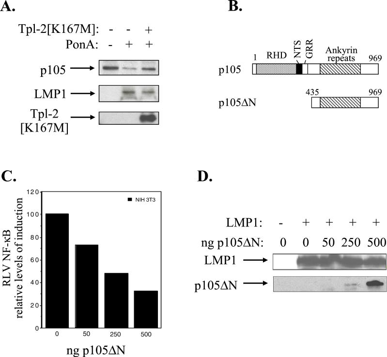
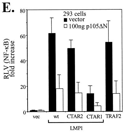
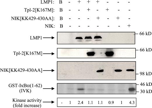
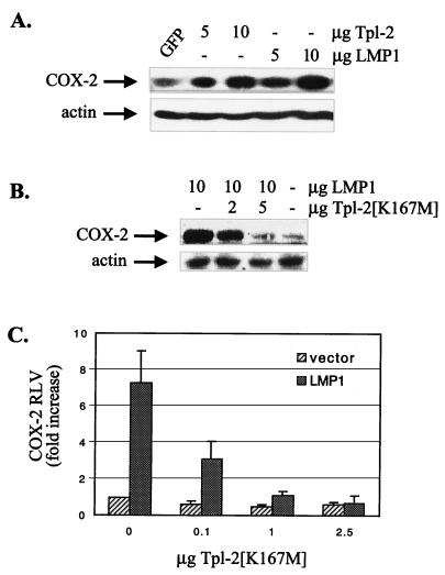
The Epstein-Barr virus-encoded latent infection membrane protein 1 (LMP1) is a pleiotropic protein, the activities of which include effects on cell transformation and phenotype, growth, and survival. The ability of LMP1 to mediate at least some of these phenomena could be attributed to the activation of the transcription factor NF-kappaB. LMP1 promotes NF-kappaB activation through the recruitment of the adapter protein TRAF2 and the formation of a dynamic multiprotein complex that includes the NF-kappaB kinase, the IkappaB kinases, and their downstream targets, IkappaBs and p105. In this study, we have identified the oncogenic kinase Tpl-2/Cot as a novel component of LMP1-induced NF-kappaB signaling. We show that Tpl-2 is expressed in primary biopsies from patients with nasopharyngeal carcinoma and Hodgkin's disease, where LMP1 is also found. Inducible expression of LMP1 promotes the activation of Tpl-2, and a catalytically inactive Tpl-2 mutant suppresses LMP1-induced NF-kappaB signaling. In colocalization and coimmunoprecipitation experiments, Tpl-2 and TRAF2 were found to interact with Tpl-2 functioning downstream of TRAF2. Consistent with this observation, catalytically inactive Tpl-2 also blocked CD40-mediated NF-kappaB activation, which largely depends on TRAF2. The ability of Tpl-2 to influence LMP1-induced NF-kappaB occurs through modulation of both IkappaBalpha and p105 functions. Furthermore, Tpl-2 was found to influence the expression of angiogenic mediators, such as COX-2 in LMP1-transfected cells. These data identify Tpl-2 as a component of LMP1 signaling downstream of TRAF2 and as a modulator of LMP1-mediated effects.


Figures

FIG. 1.
FIG. 1.
Tpl-2/Cot is detected in EBV-associated malignancies where LMP1 is commonly expressed. Shown is representative immunohistochemical staining of sections from patients with HD (A) and undifferentiated NPC (B), illustrating cytoplasmic localization of Tpl-2/Cot. Both sections shown were positive for LMP1.
FIG. 2.
FIG. 2.
LMP1 promotes the activation of Tpl-2. (A) Induction of LMP1 in 293EcR/LMP1 cells carrying an ecdysone-regulatable LMP1, following addition of a 10 μM concentration of the ecdysone analogue ponasterone A (PonA; upper panel). Lysates from 293EcR/LMP1 cells treated with PonA or from untreated cultures were analyzed for endogenous Tpl-2 levels by using the anti-Tpl-2 polyclonal antibody M20 or for β-actin levels, as indicated. Nuclear extracts from parallel cultures were examined for NF-κB DNA binding activity by EMSA (lower panel). (B) HEK 293EcR/LMP1 cells were transfected with 0.3 μg of myc-tagged Tpl-2 per 106 cells and 24 h later were analyzed for Tpl-2 expression by immunoblotting, using the M20 anti-Tpl-2 polyclonal antibody (lane 2). Lysates from untransfected cultures (lane 1) were used to verify that, under these conditions, myc-Tpl-2 is expressed at near-physiological levels. (C) LMP1 promotes the activation of Tpl-2, as measured by enhanced autophosphorylation. HEK 293EcR/LMP1 cells were transfected with 0.3 μg of myc-tagged Tpl-2 (lanes 1 to 5) or its catalytically inactive mutant, Tpl-2[K167M] (lanes 6 to 10), and then stimulated with ponasterone A for 0, 3, 4.5, 6, or 7.5 h. Duplicate cultures from each time point were pulled together to minimize differences in transfection efficiencies and processed for in vitro kinase assays as described in Materials and Methods. Parallel anti-myc immunoprecipitates were probed for Tpl-2 using the M20 anti-Tpl-2 polyclonal antibody (middle panel). Relative levels of autophosphorylation were measured on a phosphorimager and are shown in the lower panel. (D) Parental HEK 293EcR cells were transiently transfected with 0.3 μg of myc-tagged Tpl-2 and then stimulated with 10 μM ponasterone A for 0, 4.5, or 7.5 h. Tpl-2 autophosphorylation was examined as described for panel C (upper panel). Anti-myc immunoprecipitates were also probed with the anti-Tpl-2 polyclonal antibody M20 (lower panel).
FIG. 3.
FIG. 3.
Tpl-2 modulates LMP1-induced NF-κB activation. (A) Wild-type Tpl-2 induces levels of NF-κB transactivation similar to that of NIK. NIH 3T3 cells were transfected with a β-galactosidase-expressing plasmid and an NF-κB reporter in the presence of increasing amounts of NIK or Tpl-2 and 36 h later were analyzed for luciferase and β-galactosidase activities. Relative NF-κB activation (ratio of luciferase versus β-galactosidase activities; RLV) from two independent experiments is shown. Asterisks represent individual mean values of duplicate determinations from these experiments. (B) Kinase-inactive Tpl-2 inhibits LMP1-induced NF-κB binding activity in 293 cells, as determined by EMSAs (upper panel). The NF-κB complex shown contains p50-p65 heterodimers (data not shown). Nuclear extracts were also analyzed for Sp1 binding activity as a control (lower panel). (C) Kinase-inactive Tpl-2 inhibits LMP1-induced NF-κB transactivation. NIH 3T3 or HEK 293 cells were cotransfected with a galactosidase-expressing plasmid and an NF-κB reporter and 1 μg of pSG5-LMP1, in the presence of equivalent amounts of Tpl-2[K167M]. Relative NF-κB activation (ratio of luciferase versus β-galactosidase activities) (± standard deviation [SD]) from three independent experiments is shown. (D) Kinase-inactive Tpl-2 inhibits LMP1-, CTAR1 [LMP1Δ(332-386)]-, and CTAR2 (LMP1AxAxA)-induced NF-κB transactivation to similar extents. HEK 293 cells were transfected with 1 μg of pSG5-based vectors in the presence of reporter constructs and increasing amounts of Tpl-2[K167M]. To allow comparison of the inhibitory effect of kinase-inactive Tpl-2, RLVs have been normalized to 100 for each of the wild-type, CTAR1, and CTAR2 effects. In these experiments, the mean NF-κB activation values from the above LMP1 molecules were 60 (±5.4), 48.4 (±6.2), and 13 (±4). (E) Kinase-inactive GCKR does not affect LMP1-induced NF-κB transactivation. HEK 293 cells were transfected with reporter constructs and 1 μg of pSG5-LMP1, in the presence or absence of increasing amounts (0.25 or 0.5 μg) of dominant negative GCKR (GCKRdn). RLV was assessed as described for panel C. (F) Kinase-inactive Tpl-2 does not affect Cdc42-mediated NF-κB induction. HEK 293 cells were transfected with reporter constructs and 5 μg of pcDNA3-Cdc42, in the presence or absence of increasing amounts (0.25 or 0.5 μg) of dominant negative Tpl-2. RLV was assessed as described for panel C.
FIG. 4.
FIG. 4.
Tpl-2 is not a component of LMP1-induced Cdc42 activation. Serum-starved 3T3 fibroblasts were microinjected with pSG5-LMP1 (B and E) or control vector (A and D), in the presence or absence of myc-tagged Tpl-2[K167M] (C and F), as described in Materials and Methods. Phalloidin staining revealed formation of filopodia extensions in LMP1-expressing cells (B), which was not affected by the presence of kinase-inactive Tpl-2 (C). Note also the extensive formation of stress fibers in these microinjected cells. Parallel experiments using dominant negative Cdc42 verified inhibition of these LMP1-mediated cytoskeletal changes (data not shown), in agreement with a previous report (41). Transfection of the pSG5 vector alone had no effect on actin reorganization (A). Immunostaining for the p65 subunit of NF-κB (D, E, and F) revealed p65 translocation to the nucleus of LMP1-expressing cells (E) but predominantly cytoplasmic staining in control cells or cells coinjected with LMP1 and Tpl-2[K167M] (panels D and F, respectively).
FIG. 5.
FIG. 5.
Tpl-2 functions downstream of TRAF2 in LMP1 signaling. (A) Kinase-inactive Tpl-2 inhibits TRAF2-mediated NF-κB activation in a concentration-dependent manner. HEK 293 cells were transfected with 1 μg of CMV-TRAF2 and increasing amounts of Tpl-2[K167M], and RLVs were determined as described in the legend for Fig. 3C. (B) TRAF2 colocalizes with endogenous Tpl-2. BJAB cells were transfected with FLAG-tagged TRAF2 and double-stained for TRAF2 using the anti-FLAG MAb M2 and for Tpl-2 using the anti-Tpl-2 polyclonal antibody M20. Green fluorescence (a and d) identifies FLAG-TRAF2 staining in two representative examples (a to c and d to f, respectively), red fluorescence (b and e) represents Tpl-2 staining, and yellow color (c and f) corresponds to the overlay of green and red fluorescence. (C) Tpl-2 coimmunoprecipitates with TRAF2. HEK 293 cells were transfected with 5 μg of CMV-TRAF2 in the presence of equivalent amounts of myc-tagged N17Cdc42, Tpl-2, or LMP1. Following immunoprecipitation (IP) with an anti-myc tag MAb (9E10), precipitates were analyzed for TRAF2 (upper panel) or myc expression (lower panel) by immunoblotting (IB). Total lysates (8%) were also analyzed to verify the migration of TRAF2 (upper panel). Arrowheads in the lower panel show the migration of immunoprecipitated N17Cdc42, Tpl-2, and LMP1 blotted with the anti-myc tag 9E10 MAb. (D) Kinase-inactive Tpl-2 inhibits CD40-mediated NF-κB activation. HEK 293 cells were transfected with 1 μg of pcDNA3-CD40 and increasing concentrations of Tpl-2[K167M], in the presence of a luciferase and β-galactosidase plasmid, and reporter activity was determined. Mean values (± SD) from three independent experiments are shown.
FIG. 5.
FIG. 5.
Tpl-2 functions downstream of TRAF2 in LMP1 signaling. (A) Kinase-inactive Tpl-2 inhibits TRAF2-mediated NF-κB activation in a concentration-dependent manner. HEK 293 cells were transfected with 1 μg of CMV-TRAF2 and increasing amounts of Tpl-2[K167M], and RLVs were determined as described in the legend for Fig. 3C. (B) TRAF2 colocalizes with endogenous Tpl-2. BJAB cells were transfected with FLAG-tagged TRAF2 and double-stained for TRAF2 using the anti-FLAG MAb M2 and for Tpl-2 using the anti-Tpl-2 polyclonal antibody M20. Green fluorescence (a and d) identifies FLAG-TRAF2 staining in two representative examples (a to c and d to f, respectively), red fluorescence (b and e) represents Tpl-2 staining, and yellow color (c and f) corresponds to the overlay of green and red fluorescence. (C) Tpl-2 coimmunoprecipitates with TRAF2. HEK 293 cells were transfected with 5 μg of CMV-TRAF2 in the presence of equivalent amounts of myc-tagged N17Cdc42, Tpl-2, or LMP1. Following immunoprecipitation (IP) with an anti-myc tag MAb (9E10), precipitates were analyzed for TRAF2 (upper panel) or myc expression (lower panel) by immunoblotting (IB). Total lysates (8%) were also analyzed to verify the migration of TRAF2 (upper panel). Arrowheads in the lower panel show the migration of immunoprecipitated N17Cdc42, Tpl-2, and LMP1 blotted with the anti-myc tag 9E10 MAb. (D) Kinase-inactive Tpl-2 inhibits CD40-mediated NF-κB activation. HEK 293 cells were transfected with 1 μg of pcDNA3-CD40 and increasing concentrations of Tpl-2[K167M], in the presence of a luciferase and β-galactosidase plasmid, and reporter activity was determined. Mean values (± SD) from three independent experiments are shown.
FIG. 6.
FIG. 6.
LMP1-induced Tpl-2-mediated p105 degradation contributes to NF-κB transactivation. (A) Kinase-inactive Tpl-2 inhibits LMP1-mediated p105 degradation. HA-tagged Tpl-2[K167M] (1 μg) and 3 μg of myc-tagged p105 were transfected in 293EcR/LMP1 cells. Following treatment with the ecdysone analogue ponasterone A for 9 h, cell lysates were isolated and analyzed for LMP1, Tpl-2, or myc-p105 expression by immunoblotting. (B) Structure of p105 and the N-terminus-deleted, nondegradable mutant used in this study. RHD, rel-homology domain; NTS, nuclear translocation signal; GRR, glycine-rich region. Both constructs carry a myc tag in their N termini. (C) Transfection of p105ΔN in NIH 3T3 cells inhibits LMP1-induced NF-κB activation. The relative decrease in NF-κB luciferase values (RLVs) is shown in histogram form, in which LMP1 has been given the arbitrary value of 100. The actual increase in NF-κB transactivation induced by LMP1 in this representative assay was 8.5-fold. (D) Identical lysates from the experiment shown in panel C were analyzed for LMP1 expression in order to verify that the inhibitory effects of p105ΔN on LMP1-induced NF-κB activation were not due to altered LMP1 levels (upper panel). Deleted p105 expression was assessed by immunoblot analysis using anti-myc tag 9E10 MAb (lower panel). (E) Transfection of a low plasmid concentration (100 ng) of p105ΔN significantly inhibits NF-κB transactivation induced by 1 μg of LMP1, LMP1 CTAR1 [LMP1Δ(332-386)], or LMP1 CTAR2 (LMP1AxAxA) in HEK 293 cells. The effects of this deleted p105 mutant on TRAF2-mediated NF-κB reporter activity are also shown. Data (RLVs) are the mean values (± SD) from three independent experiments.
FIG. 6.
FIG. 6.
LMP1-induced Tpl-2-mediated p105 degradation contributes to NF-κB transactivation. (A) Kinase-inactive Tpl-2 inhibits LMP1-mediated p105 degradation. HA-tagged Tpl-2[K167M] (1 μg) and 3 μg of myc-tagged p105 were transfected in 293EcR/LMP1 cells. Following treatment with the ecdysone analogue ponasterone A for 9 h, cell lysates were isolated and analyzed for LMP1, Tpl-2, or myc-p105 expression by immunoblotting. (B) Structure of p105 and the N-terminus-deleted, nondegradable mutant used in this study. RHD, rel-homology domain; NTS, nuclear translocation signal; GRR, glycine-rich region. Both constructs carry a myc tag in their N termini. (C) Transfection of p105ΔN in NIH 3T3 cells inhibits LMP1-induced NF-κB activation. The relative decrease in NF-κB luciferase values (RLVs) is shown in histogram form, in which LMP1 has been given the arbitrary value of 100. The actual increase in NF-κB transactivation induced by LMP1 in this representative assay was 8.5-fold. (D) Identical lysates from the experiment shown in panel C were analyzed for LMP1 expression in order to verify that the inhibitory effects of p105ΔN on LMP1-induced NF-κB activation were not due to altered LMP1 levels (upper panel). Deleted p105 expression was assessed by immunoblot analysis using anti-myc tag 9E10 MAb (lower panel). (E) Transfection of a low plasmid concentration (100 ng) of p105ΔN significantly inhibits NF-κB transactivation induced by 1 μg of LMP1, LMP1 CTAR1 [LMP1Δ(332-386)], or LMP1 CTAR2 (LMP1AxAxA) in HEK 293 cells. The effects of this deleted p105 mutant on TRAF2-mediated NF-κB reporter activity are also shown. Data (RLVs) are the mean values (± SD) from three independent experiments.
FIG. 7.
FIG. 7.
Involvement of Tpl-2 in LMP1-mediated IκBα activation. HEK 293 cells were transiently transfected with 2.5 μg of pSG5-LMP1 or control vector in the presence or absence of equal amounts of kinase-inactive Tpl-2 (lanes 4 and 6) or NIK (lanes 5 and 7). Lysates were subjected to immunoblot analysis for LMP1, Tpl-2, or NIK expression (upper three panels). Endogenous IKKα was immunoprecipitated using the H744 polyclonal anti-IKKα antibody (Santa Cruz Biotechnology) and analyzed for kinase activity in an in vitro kinase assay (IVK) using GST-IκBα(1-62) as the substrate. As a positive control, lysates from wild-type NIK-transfected cells were used (lane 8). IκBα phosphorylation was measured on a phosphorimager, and relative levels of kinase activity are shown. In three independent experiments, LMP1 induced a mean of 2.1 (±0.4)-fold increase in IκBα activation, while kinase-inactive Tpl-2 consistently suppressed this effect by more than 85%. Background phosphorylation (B, lane 1) from mock immunoprecipitation (in the absence of IKKα antibody) is also shown. Expression of wild-type Tpl-2 has been previously shown to induce IκBα phosphorylation in 293 cells (33).
FIG. 8.
FIG. 8.
Tpl-2 modulates the expression of the angiogenic factor COX-2. (A) LMP1 and Tpl-2 expression in HEK 293 cells transduces signals leading to COX-2 up-regulation. Cells were transfected with increasing amounts of LMP1, Tpl-2, or GFP-expressing vectors as indicated and 36 h later total lysates were analyzed for COX-2 or β-actin expression by immunoblotting. (B) Catalytically inactive Tpl-2 inhibits the ability of LMP1 to induce COX-2 expression. HEK 293 cells were transfected with 10 μg of LMP1 in the absence or presence of increasing amounts of Tpl-2[K167M] as indicated, and 36 h later total lysates were analyzed for COX-2 or β-actin expression by immunoblotting. (C) Tpl-2[K167M] suppresses LMP1-induced COX-2 promoter activity. HEK293 cells were cotransfected with 0.6 μg of COX-2 promoter-luciferase reporter construct, 0.2 μg of Renilla plasmid, 2.5 μg of pSG5-LMP1, and increasing amounts of Tpl-2[K167M], as indicated. The fold increase in RLVs (ratio of luciferase versus Renilla values) is shown.

References

    1. Belich, M. P., A. Salmeron, L. H. Johnston, and S. C. Ley. 1999. Tpl-2 kinase regulates the proteolysis of the NF-κB-inhibitory protein NF-κB1 p105. Nature 397:363-368. - PubMed
    1. Cahir-McFarland, E. D., D. M. Davidson, S. L. Schauer, J. Duong, and E. Kieff. 2000. NF-κB inhibition causes spontaneous apoptosis in Epstein-Barr virus-transformed lymphoblastoid cell lines. Proc. Natl. Acad. Sci. USA 97:6055-6060. - PMC - PubMed
    1. Cahir-McFarland, E. D., K. M. Izumi, and G. Mosialos. 1999. Epstein-Barr virus transformation: involvement of latent membrane protein 1-mediated activation of NF-κB. Oncogene 18:6959-6964. - PubMed
    1. Cammarano, M. S., and A. Minden. 2001. Dbl and the Rho GTPases activate NF-κB by IκB kinase (IKK)-dependent and IKK-independent pathways. J. Biol. Chem. 276:25876-25882. - PubMed
    1. Ceci, J. D., C. P. Patriotis, C. Tsatsanis, A. M. Makris, R. Kovatch, D. A. Swing, N. A. Jenkins, P. N. Tsichlis, and N. G. Copeland. 1997. Tpl-2 is an oncogenic kinase that is activated by carboxy-terminal truncation. Genes Dev. 11:688-700. - PubMed

Publication types

MeSH terms

Substances

LinkOut - more resources