. 2002 May 3;296(5569):922-7.
doi: 10.1126/science.1069398. Epub 2002 Apr 4.

Genomic instability in mice lacking histone H2AX

Affiliations

Genomic instability in mice lacking histone H2AX

Arkady Celeste et al. Science. .

Abstract

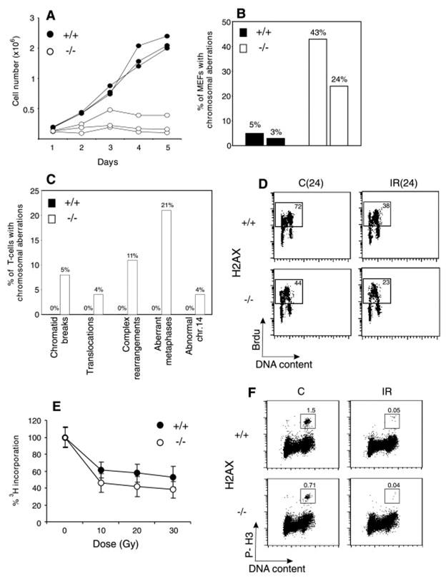
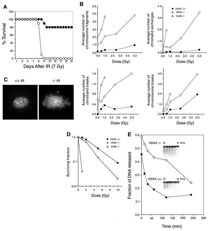
Higher order chromatin structure presents a barrier to the recognition and repair of DNA damage. Double-strand breaks (DSBs) induce histone H2AX phosphorylation, which is associated with the recruitment of repair factors to damaged DNA. To help clarify the physiological role of H2AX, we targeted H2AX in mice. Although H2AX is not essential for irradiation-induced cell-cycle checkpoints, H2AX-/- mice were radiation sensitive, growth retarded, and immune deficient, and mutant males were infertile. These pleiotropic phenotypes were associated with chromosomal instability, repair defects, and impaired recruitment of Nbs1, 53bp1, and Brca1, but not Rad51, to irradiation-induced foci. Thus, H2AX is critical for facilitating the assembly of specific DNA-repair complexes on damaged DNA.


Figures

Fig. 1
Fig. 1. H2AX deficiency results in growth retardation and genomic instability, but does not abrogate irradiation-induced cell-cycle checkpoints. (A) Growth kinetics of three independent H2AX−/− (○) and wild-type (●) MEFs at passage 1
(B) Percentage of cells with chromosome aberrations from two different H2AX wild-type (filled bars) and knockout (open bars) MEFs. (C). Aberrations found in wild-type (+/+) (left bars) and knockout (−/−) (right bars) activated T cells. (D) Irradiation-induced G1 to S checkpoint. Exponentially growing passage 1 MEFs were untreated (C) or irradiated (IR) with 10 Gy of γ-irradiation, then grown for 24 hours in the presence of BrdU. The percentage of cycling (BrdU+) cells is indicated. (E) Irradiation-induced S-phase checkpoint. The [3H]thymidine incorporation in unirradiated cultures was set to 100%. (F) Irradiation-induced G2-M checkpoint. Cells were either untreated or irradiated with 10 Gy, then incubated for 1 hour at 37°, and cells in mitosis were identified by costaining with PI and antibody to phospho-histone H3 (P-H3). The percentage of cells in mitosis is indicated.
Fig. 2
Fig. 2. H2AX−/− mice are radiation sensitive and immortalized MEFs exhibit defective DNA repair
(A) Survival of 4-week-old H2AX−/− and littermate controls exposed to 7 Gy of whole-body γ-irradiation. Ten H2AX−/− (○) and 10 control mice (● 5 H2AX+/− and 5 H2AX+/+ mice) were used. (B) Average number of chromosomal aberrations per metaphase (breaks, fragments, and exchanges) induced by IR in H2AX+/+, H2AX−/−, and Ku80−/− immortalized MEFs. At IR doses exceeding 1 Gy, Ku80−/− metaphases exhibited massive chromosomal fragmentation. At least 20 metaphases were examined for each genotype. (C) DAPI (4′,6-diamidino-2-phenylindole) staining of H2AX+/+ and H2AX−/− fibroblast nuclei 24 hours after treatment with 10-Gy γ-irradiation. Ten percent of H2AX−/− cells (n = 400) and 0.25% of wild-type nuclei (n = 400) showed extensive nuclear fragmentation. (D) Radiation sensitivity of H2AX+/+ (●), H2AX−/− (○), and Ku80−/− (Δ) fibroblasts, plotted as the fraction of surviving cells relative to unirradiated samples of the same genotype. (E) Rejoining of DNA DSBs produced by 80-Gy γ-irradiation. (Inset) The fraction of DNA released into the well is plotted.
Fig. 3
Fig. 3. Impaired immunoglobulin class-switch recombination in H2AX−/− mice
(A) (Upper panel) Two-color flow cytometric analysis of IgG1 expression on CFSE-labeled B cells that were stimulated with LPS plus IL-4 for 4 days. The percentage of cells expressing IgG1 that had undergone a given number of cell divisions is quantified in the lower panel. (B) Sera from 6-week-old H2AX−/− (○) and age-matched wild-type (●) mice were collected, and total IgM, IgG1, IgA, and IgG3 levels were determined by enzyme-linked immunosorbent assay. (C) Mice were immunized with TNP-KLH, and anti-TNP–specific IgM or IgG1 serum levels were measured at 0, 5, 12, and 21 days after immunization. Data are plotted as the average dilution (mean ± SD) determined in five mice of each genotype.
Fig. 4
Fig. 4. Defective spermatogenesis in H2AX−/− mice
(A) Comparison of testis size in 2-month-old H2AX wild-type (+/+) and knockout (−/−) mice. Bar, 2 mm. (B) Sections of seminiferous tubules of 7-week-old (+/+ and −/−) littermates stained with hematoxylin-eosin. Magnification, ×10. (C) Hematoxylin-eosin-stained sections of epididymis from the same mice shown in (B). Magnification, ×40. (D) High-magnification (×100) images of periodic acid–Schiff–stained paraffin sections of seminiferous tubules of 2-month old wild-type (+/+) (left panel) and knockout (−/−) (two right panels) mice. Primary spermatocytes in early pachytene (EP), late pachytene (LP), and zygotype (Z) are indicated. Apoptotic nuclei with condensing chromatin are present in H2AX−/− tubules (arrows). (E) TUNEL (terminal deoxynucleotidyl transferase–mediated dUTP nick-end labeling) assay detects very few apoptotic cells in normal tubules (arrows, left panel), whereas H2AX-mutant tubules contain a large number of dying cells. Magnification, ×40. (F to I) Indirect immunofluorescence of H2AX+/− (+/−; left panels) and H2AX−/− (−/−; right panels) pachytene spermatocytes. (F) Merged image of Scp3 (red) and γ-H2AX (green). (G) Merged image of Scp3 (green) and Scp1 (red). (H) Merged image of Scp1 (red) and Mlh1 (green). (I) Diplotene (left) and diakinesis (right) H2AX+/− stages (two individual cells separated by the dotted line), and fragmented synaptonemal complex in H2AX−/− spermatocyte visualized with antibody to Scp3 (red) and counterstained with DAPI (blue). For (F) to (H), the arrowhead indicates the Y chromosome, and the arrow shows the X chromosome. Bar [(F) to (I)], 10 μm.
Fig. 5
Fig. 5. DNA-repair foci formation in B cells and fibroblasts
H2AX wild-type (+/+) and knockout (−/−) B cells were stimulated for 2 days with LPS and IL-4, left untreated (C) or exposed to 500 cGy of γ-irradiation (IR), and then incubated at 37°C before staining for (A) Nbs1, (B) 53bp1, (C) Brca1, or (D) Rad51. Because irradiation-induced Nbs1 foci assemble after the appearance of 53bp1, Brca1, and Rad51 foci, recovery after irradiation was 8 hours for Nbs1 and 4 hours for 53bp1, Brca1, and Rad51. Similar results were obtained at 0.75 hours, 2 hours, and 4 hours after irradiation (9). Of irradiated wild-type cells, 57% contained Rad51 foci, 74% of which had more than three foci per cell; of irradiated H2AX−/− cells, 53% contained Rad51 foci, 72% of which had more than three foci per cell. (E). Nbs1 (green) and PCNA (red) staining in detergent-extracted wild-type and H2AX−/− fibroblasts. Images were merged to determine colocalization. PCNA/Nbs1 clusters are localized to heterochromatic regions coincident with intense DAPI staining (blue).

References

    1. Redon C, et al. Curr Opin Genet Dev. 2002;12:162. - PubMed
    1. Rogakou EP, Pilch DR, Orr AH, Ivanova VS, Bonner WM. J Biol Chem. 1998;273:5858. - PubMed
    1. Rogakou EP, Boon C, Redon C, Bonner WM. J Cell Biol. 1999;146:905. - PMC - PubMed
    1. Chen HT, et al. Science. 2000;290:1962. - PMC - PubMed
    1. Petersen S, et al. Nature. 2001;414:660. - PMC - PubMed

Publication types

MeSH terms