. 2002 Apr 1;16(7):796-805.
doi: 10.1101/gad.972702.

A molecular link between gene-specific and chromosome-wide transcriptional repression

Affiliations

A molecular link between gene-specific and chromosome-wide transcriptional repression

Diana S Chu et al. Genes Dev. .

Abstract

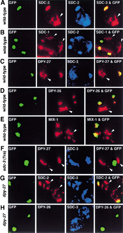
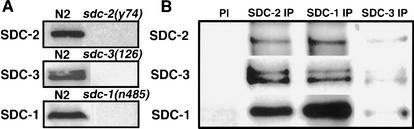
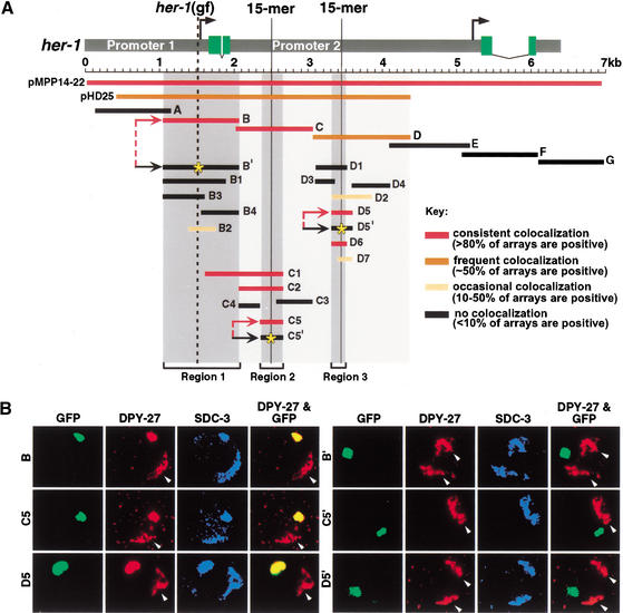
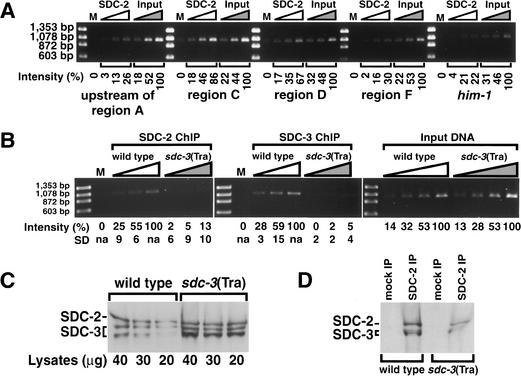
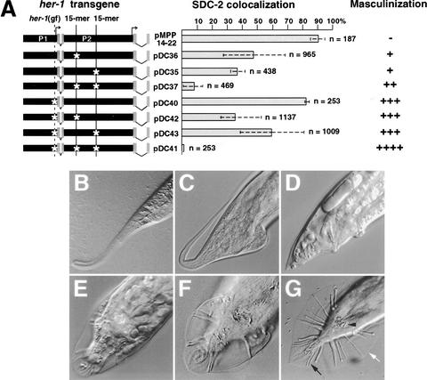
Gene-specific and chromosome-wide mechanisms of transcriptional regulation control development in multicellular organisms. SDC-2, the determinant of hermaphrodite fate in Caenorhabditis elegans, is a paradigm for both modes of regulation. SDC-2 represses transcription of X chromosomes to achieve dosage compensation, and it also represses the male sex-determination gene her-1 to elicit hermaphrodite differentiation. We show here that SDC-2 recruits the entire dosage compensation complex to her-1, directing this X-chromosome repression machinery to silence an individual, autosomal gene. Functional dissection of her-1 in vivo revealed DNA recognition elements required for SDC-2 binding, recruitment of the dosage compensation complex, and transcriptional repression. Elements within her-1 differed in location, sequence, and strength of repression, implying that the dosage compensation complex may regulate transcription along the X chromosome using diverse recognition elements that play distinct roles in repression.


Figures

Figure 1
Figure 1
The X-chromosome dosage compensation machinery localizes to her-1 regulatory regions in vivo. Confocal images of an individual gut nucleus (AF) or an embryonic nucleus (G,H) from wild-type or mutant [sdc-3(Tra) or dpy-27] XX animals immunostained with SDC, DPY, or MIX antibodies, as indicated in each panel. The nuclei contain extrachromosomal DNA arrays carrying multiple copies of her-1 regulatory regions (plasmid pHD25 of Fig. 3A), lac operator repeats (lacO), and a transgene encoding a LacI–GFP fusion protein. LacI–GFP repressor binding to lacO permits array detection by GFP autofluorescence. Colocalization (yellow) between arrays (green) and antibodies (red) in the merged images (right panels) showed association of the protein with her-1 regulatory sequences. Arrowheads mark the X chromosomes. Consistent with sdc-3(Tra) causing derepression of her-1, it blocks SDC and DPY proteins from associating with her-1.
Figure 2
Figure 2
The SDC proteins form a complex in vivo. (A) Detection of SDC proteins in embryonic extracts. Western blots of extracts from wild-type (N2) or sdc (null) mutant embryos carrying a deletion or nonsense mutation in the sdc gene were probed with the SDC antibody indicated on the left. Proteins of 250 kD, 240 kD (a doublet), and 140 kD were detected by SDC-2, SDC-3, or SDC-1 antibodies, respectively, in wild-type but not sdc (null) extracts, showing antibody specificity. (B) Antibody to any one SDC protein coimmunoprecipitated all three SDC proteins. Coimmunoprecipitations were performed with each SDC antibody on wild-type embryonic extracts. The coimmunoprecipitated material was separated by SDS-PAGE and immunoblotted with the antibody indicated on the left. (PI) Preimmune sera for SDC-1 antibody, (IP) immunoprecipitation.
Figure 3
Figure 3
SDC-2 localization to her-1 is specified by three distinct DNA recognition elements whose binding capacity is disrupted by specific mutations. (A) Schematic of the her-1 gene and summary of subregions tested for SDC-2 colocalization by the array assay. Transcription from the P1 promoter produces the functional male-specific her-1 transcript, including four exons (green). A promoter (P2) resides within the second intron of her-1. P2 is coregulated with P1 and makes a 0.8-kb transcript of unknown function that includes the last two exons of her-1 (Trent et al. 1991; Perry et al. 1993). The degree of SDC-2 colocalization with her-1 regions is shown by color, with the key on the right. The three smallest regions with strong SDC-2 colocalization (region 1 [B], region 2 [C5], and region 3 [D6]) are shown by dark gray shading. C5 and D6 share an identical 15-bp element (solid vertical lines) and 50% overall sequence identity. B has no obvious similarity with C5 or D5 but contains the site of her-1(gf) (dashed vertical line). SDC-2 colocalization was completely disrupted by a randomized 15-mer in either C5 or D5 and by the G → A transition of her-1(gf) in B (yellow stars in B‘, C5′, and D5′). (B) Confocal images of an individual gut nucleus from a wild-type XX animal bearing GFP-tagged extrachromosomal arrays (green) with either wild-type (B, C5, and D5) or mutant (B‘, C5′, and D5′) versions of regions 1–3. Animals were immunostained with antibodies to DPY-27 (red) or SDC-3 (blue). Colocalization between the array and protein appears as yellow in the merged image. Arrowheads indicate X chromosomes.
Figure 4
Figure 4
Chromatin immunoprecipitation experiments (ChIP) show the interaction of SDC proteins with the endogenous her-1 gene. (A) PCR analysis of DNA from a ChIP performed with SDC-2 antibodies and lysates of formaldehyde cross-linked XX embryos. Primer sets specific to her-1 regions or a control gene (him-1) were used for PCRs with mock-precipitated DNA (M) and twofold serial dilutions of SDC-2-precipitated DNA (SDC-2) or input DNA (Input). The intensity of the PCR band produced by each primer pair from IP-enriched DNA was normalized to the corresponding PCR band produced from the highest concentration of input DNA. Regions D and C of her-1 were specifically enriched above him-1 control DNA by threefold or fourfold, respectively. (Primers flanking him-1 produced a PCR product from IP-enriched DNA of 22% normalized intensity, whereas primers flanking regions D and C produced bands of 67% and 86%, respectively, normalized intensity.) (B) PCR analysis with region C primers was performed on twofold serial dilutions of DNA from a ChIP using SDC-2 or SDC-3 antibodies and lysates of formaldehyde-treated wild-type or sdc-3(Tra) XX embryos. The intensity of each PCR band was normalized to the intensity of the PCR band made from the highest concentration of IP-enriched DNA from the wild-type lysate. The average intensities and standard deviations were calculated from four sets of PCR analyses on material from two independent ChIP experiments. The specificity of the ChIP protocol was shown by the precipitation of region C DNA from wild-type but not mutant lysates. Similar levels of region C DNA were detected by PCR using twofold serial dilutions of wild-type and sdc-3(Tra) input lysates. (C,D) Similar levels of SDC-2 and SDC-3 were detected by Western blot analysis of either (C) whole lysates or (D) SDC-2 IP material from lysates of formaldehyde-treated wild-type and sdc-3(Tra) embryos.
Figure 5
Figure 5
The relative contributions of the three her-1 recognition elements differ for SDC binding and her-1 repression. (A) Mutation of specific DNA sequences within full-length her-1 transgenes disrupts SDC-2 localization to her-1 and repression of her-1. her-1 constructs included in transgenic arrays are depicted by diagrams on the left, with mutations indicated by white stars. The percent SDC-2 localization to transgenic arrays is represented by gray bars. The standard deviation between lines assayed is represented by a dotted line. (n) Total number of nuclei scored in all lines. Masculinization caused by the full-length her-1 transgenes was quantified (see Materials and Methods) and then rated as (−) none, (+) weak, (++) moderate, (+++) strong, or (++++) severe. (BG) Examples of masculinization caused by derepression of her-1. DIC photomicrographs of tails from (B) a wild-type XX hermaphrodite, (CF) XX animals masculinized by full-length her-1 transgenes, and (G) a wild-type XO male. Lateral views (BD) and ventral views (EG). (White arrow) male fan; (black arrow) male sensory rays; (black arrowhead) male spicules.

References

    1. Aparicio OM, Billington BL, Gottschling DE. Modifiers of position effect are shared between telomeric and silent mating-type loci in S. cerevisiae. Cell. 1991;66:1279–1287. - PubMed
    1. Carmi I, Kopczynski JB, Meyer BJ. The nuclear hormone receptor SEX-1 is an X-chromosome signal that determines nematode sex. Nature. 1998;396:168–173. - PubMed
    1. Chuang P-T, Lieb JD, Meyer BJ. Sex-specific assembly of a dosage compensation complex on the nematode X chromosome. Science. 1996;274:1736–1739. - PubMed
    1. Davis TL, Meyer BJ. SDC-3 coordinates the assembly of a dosage compensation complex on the nematode X chromosome. Development. 1997;124:1019–1031. - PubMed
    1. Dawes HE, Berlin DS, Lapidus DM, Nusbaum C, Davis TL, Meyer BJ. Dosage compensation proteins targeted to X chromosomes by a determinant of hermaphrodite fate. Science. 1999;284:1800–1804. - PubMed

Publication types

MeSH terms