. 2002 Apr 1;16(7):859-69.
doi: 10.1101/gad.965602.

Coordination of chondrogenesis and osteogenesis by fibroblast growth factor 18

Affiliations

Coordination of chondrogenesis and osteogenesis by fibroblast growth factor 18

Zhonghao Liu et al. Genes Dev. .

Abstract

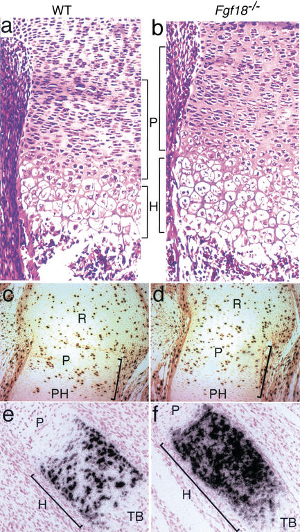
Gain of function mutations in fibroblast growth factor (FGF) receptors cause chondrodysplasia and craniosynostosis syndromes. The ligands interacting with FGF receptors (FGFRs) in developing bone have remained elusive, and the mechanisms by which FGF signaling regulates endochondral, periosteal, and intramembranous bone growth are not known. Here we show that Fgf18 is expressed in the perichondrium and that mice homozygous for a targeted disruption of Fgf18 exhibit a growth plate phenotype similar to that observed in mice lacking Fgfr3 and an ossification defect at sites that express Fgfr2. Mice lacking either Fgf18 or Fgfr3 exhibited expanded zones of proliferating and hypertrophic chondrocytes and increased chondrocyte proliferation, differentiation, and Indian hedgehog signaling. These data suggest that FGF18 acts as a physiological ligand for FGFR3. In addition, mice lacking Fgf18 display delayed ossification and decreased expression of osteogenic markers, phenotypes not seen in mice lacking Fgfr3. These data demonstrate that FGF18 signals through another FGFR to regulate osteoblast growth. Signaling to multiple FGFRs positions FGF18 to coordinate chondrogenesis in the growth plate with osteogenesis in cortical and trabecular bone.


Figures

Figure 1
Figure 1
Fgf18 expression in E14.5 developing limb. (a) Proximal humerus growth plate counterstained with hematoxylin and viewed in bright field. (b) Fgf18 in situ hybridization viewed in dark field. Note the expression of Fgf18 in the perichondrium. (c) Knee joint counterstained with hematoxylin and viewed in bright field. (d) Fgf18 in situ hybridization viewed in dark field. Note the expression of Fgf18 in the joint tissue and perichondrium. (R) Reserve chondrocytes; (P) proliferating chondrocytes; (PH) prehypertrophic chondrocytes; (Fe) femur; (Ti) tibia.
Figure 2
Figure 2
Generation of an Fgf18-LacZ targeted allele. (a) A schematic representation of the Fgf18 genomic locus, the targeting vector and the mutant allele generated following homologous recombination. (b) Southern blot identification of the Fgf18 targeted allele in embryonic stem cells using a 3′ probe and a HindIII digest. (c) Southern blot identification of the Fgf18 targeted allele in tail DNA using a 3′ probe and a HindIII digest. The 3′ probe (as indicated in a) identifies the wild-type allele as an 11-kb HindIII fragment and the mutant allele as a 4-kb HindIII fragment. (d) In situ hybridization detection of Fgf18 mRNA expression in the developing palatal shelf in wild-type mice (upper panels) and in Fgf18−/− tissue (lower panels). Brightfield and darkfield images are shown. Note the absence of expression in Fgf18−/− tissue (lower panels). (ei) Staining for β-galactosidase enzyme activity in Fgf18/−/+ cranium and limbs of E14.5 mice. (e) A sagittal section through the parietal bone showing β-galactosidase enzyme activity in the periosteum and endosteum. (f,g) Dorsal (f) and ventral (g) views of a whole mount-stained limb showing β-galactosidase enzyme activity in skeletal elements. (h, i) Histological sections through the distal limb stained for β-galactosidase enzyme activity. A planar section is shown in (h) and a cross section is shown in (i). (D) Dorsal; (V) ventral.
Figure 3
Figure 3
Morphological analysis of the skeletal phenotype in Fgf18−/− mice. (a) Alizarin red- and alcian blue-stained skeletal preparations of neonatal (P0) mice. The Fgf18−/− skeleton is shown below and a wild-type littermate is shown above. Note the deformed ribs and smaller thoracic cavity in Fgf18−/− mice. (b) Alizarin red- and alcian blue-stained skull from an E17.5 wild-type littermate (upper), an Fgf18−/− embryo (middle), and an Fgfr3−/− embryo (lower), showing decreased growth of cranial bones in the Fgf18−/− but not in the Fgfr3−/− skull. (c,d) Higher-magnification view of the hind limbs from an E17.5 Fgf18−/− embryo (c) and Fgfr3−/− embryo (d). Each panel also shows a wild-type littermate control. (*) Indicates a noticeable defect in Fgf18−/− mice. The arrow indicates the ossification zone in the metatarsal bones. (e,f) Forelimbs from P0 (e) and E17.5 (f) embryos showing delayed ossification in Fgf18−/− mice. Note that the P0 Fgf18−/− limb looks similar to the E17.5 wild-type limb. (g,h) Palate morphology from a wild-type littermate (g) and a neonatal Fgf18−/− mouse (h). Note the complete cleft palate in the Fgf18−/− mouse.
Figure 4
Figure 4
Histological analysis and identification of proliferating and hypertrophic chondrocytes using BrdU immunohistochemistry and type X collagen in situ hybridization. (a,b) Hematoxylin and eosin-stained section of the distal femur from an E16.5 wild-type embryo (a) and an Fgf18−/− embryo (b). Note the increased height of the Fgf18−/− proliferating and hypertrophic zones relative to that of the control. (c,d) Immunohistochemical detection of BrdU-labeled chondrocytes in the epiphyseal growth plate of the distal femur from an E16.5 wild-type embryo (c) and an Fgf18−/− embryo (d). (e,f) Type X collagen expression in the distal fibula growth plate of an E18.5 wild-type littermate (e) and Fgf18−/− mouse (f). (R) Reserve chondrocytes; (P) proliferating chondrocytes; (PH) prehypertrophic chondrocytes; (H) hypertrophic chondrocytes; (TB) trabecular bone.
Figure 5
Figure 5
In situ detection of Ihh and patched in the developing growth plate. (ad) Ihh expression in the distal fibula growth plate of an E16.5 wild-type (a,c) and Fgf18−/− (b,d) mouse. (eh) patched expression in the distal humerus growth plate of an E16.5 wild-type (e,g) and Fgf18−/− (f,h) mouse. Brightfield (a,b,e,f) and darkfield (c,d,g,h) images are shown. (P) Proliferating chondrocytes; (PH) prehypertrophic chondrocytes; (H) hypertrophic chondrocytes.
Figure 6
Figure 6
In situ detection of osteopontin, Cbfa1, and Vegf in the developing growth plate of the humerus at E15.5. (a,b) Brightfield images. (ch) Darkfield images. (c,d) Op expression. (e,f) Cbfa1 expression. (g,h) Vegf expression. (a,c,e,g) Sections from wild-type littermates. (b,d,f,h) Sections from Fgf18−/− mice.
Figure 7
Figure 7
Model for Fgf18 regulation of long bone growth. In a linear model of chondrogenesis, chondrocytes sequentially develop through reserve (R), proliferating (P), prehypertrophic (PH), and hypertrophic (H) stages. The rectangles indicate the relative expression domains of signaling molecules in the growth plate. Previous studies show that FGFR3 inhibits (1) chondrocyte proliferation, (2) chondrocyte differentiation, and (3) Ihh expression in prehypertrophic chondrocytes (Naski et al. 1998). Fgf18 is expressed in the perichondrium and is proposed to activate FGFR3 signaling in proliferating and prehypertrophic chondrocytes (green arrow). FGF18 may also signal to FGFR1 in hypertrophic chondrocytes and FGFR2 in the perichondrium and in trabecular bone. Decreased IHH signaling through Patched (PTC) and Smoothened (SMO) results in decreased PTHrP expression in the periarticular perichondrium and signaling to the PTHrP receptor (PTHrP-R) in prehypertrophic chondrocytes. PTHrP-R signaling delays chondrocyte maturation. FGF18 could inhibit chondrocyte proliferation either directly through the action of FGFR3 in proliferating chondrocytes or indirectly by repressing the Ihh–Patched–PTHrP signaling pathway. Contrary to its function in chondrogenesis, FGF18 positively regulates osteogenesis. In a linear model of osteogenesis, mesenchymal cells (MC) develop into osteogenic progenitor cells (OP), which express Cbfa1. OP cells then differentiate into mature osteoblasts (OB) which eventually become entrapped in bone as osteocytes (OC). In cortical bone, FGF18 promotes osteoblast maturation/proliferation, but has no effect on the formation of the osteoprogenitor cell population. This suggests that FGF signaling affects differentiation of the committed osteoprogenitor cell. The delayed formation of trabecular bone suggests that FGF18 signals directly through FGFR1 or indirectly through other factors to regulate ossification of hypertrophic chondrocytes. FGF18 signaling to FGFR3 in proliferating chondrocytes and potentially to FGFR2 in the perichondrium/periosteum or FGFR1 in hypertrophic chondrocytes places FGF18 in a position to coordinate rates of growth and differentiation in developing bone.

References

    1. Alvarez J, Horton J, Sohn P, Serra R. The perichondrium plays an important role in mediating the effects of TGF-β1 on endochondral bone formation. Dev Dyn. 2001;221:311–321. - PubMed
    1. Caplan AI, Pechak DG. The cellular and molecular embryology of bone formation. In: Peck WA, editor. Bone and mineral research. New York, NY: Elsevier Science Publishers; 1987. pp. 117–183.
    1. Chen L, Li C, Qiao W, Xu X, Deng C. A Ser(365)→Cys mutation of fibroblast growth factor receptor 3 in mouse downregulates Ihh/PTHrP signals and causes severe achondroplasia. Hum Mol Genet. 2001;10:457–465. - PubMed
    1. Chen Y, Struhl G. Dual roles for patched in sequestering and transducing Hedgehog. Cell. 1996;87:553–563. - PubMed
    1. Colvin JS, Bohne BA, Harding GW, McEwen DG, Ornitz DM. Skeletal overgrowth and deafness in mice lacking fibroblast growth factor receptor 3. Nat Genet. 1996;12:390–397. - PubMed

Publication types

MeSH terms