. 2002 Apr 1;16(7):880-91.
doi: 10.1101/gad.221002.

Role of DIVARICATA in the control of dorsoventral asymmetry in Antirrhinum flowers

Affiliations

Role of DIVARICATA in the control of dorsoventral asymmetry in Antirrhinum flowers

Lisete Galego et al. Genes Dev. .

Abstract

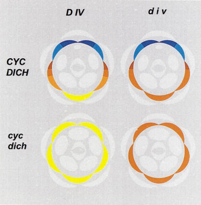
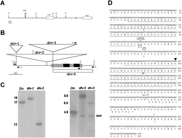
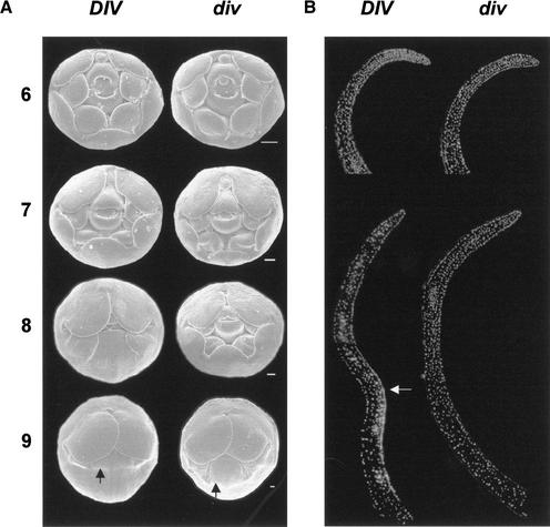
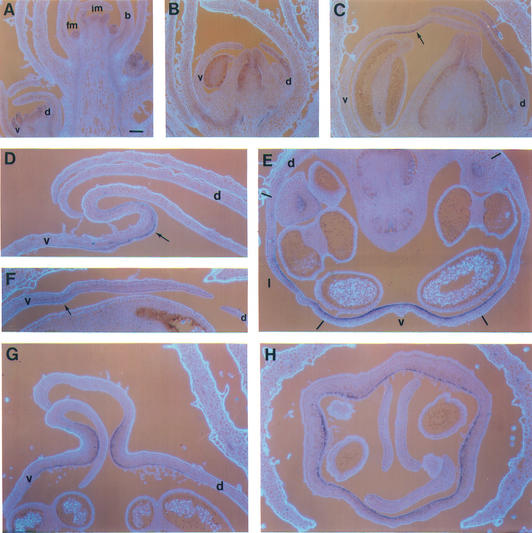
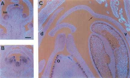
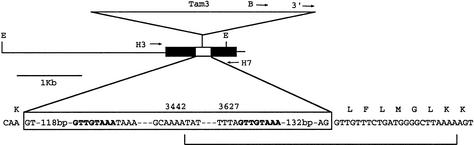
Dorsoventral asymmetry of the Antirrhinum corolla depends on expression of the CYC and DICH genes in dorsal petals. One role of these genes is to inhibit DIVARICATA (DIV), a determinant of ventral identity. Therefore, in cyc;dich double mutants ventral identity spreads all around the flower. We show that DIV encodes a protein belonging to the MYB family of transcription factors. Early on in corolla development, DIV affects specifically the growth of ventral and lateral petals but is transcribed in all petals. Analysis of a closely related gene suggests that the lack of effect on dorsal petals is not due to redundancy. More likely, therefore, DIV is regulated posttranscriptionally through a mechanism that depends on CYC and DICH. Later on, DIV affects growth and cell types and is transcribed mostly in a single layer of cells of ventral and lateral petals. This late pattern may itself depend on DIV activity because it fails to be established in a transcribed but inactive div mutant and, conversely, spreads all around the flower in cyc;dich double mutants.


Figures

Figure 1
Figure 1
Floral diagrams of wild type and dorsoventral mutants in Antirrhinum. Petals are in various colors, with all other organs in pale gray. Ventral identity is in yellow, lateral in brown, and dorsal in blue. The dorsal petal is divided in dorsal (dark blue) and lateral (pale blue) halves. In single dich mutants the dorsal half adopts a pale blue identity. The lateral petal is divided in three regions (shades of brown). div has very little or no effect on its most dorsal region (darkest brown) and affects its most ventral region (palest brown). In Div;div heterozygotes the lateral petal remains as in wild type while the ventral petal adopts the palest brown identity (see Almeida et al. 1997). Note that unlike cyc;dich, the div mutation has no effect on stamens.
Figure 2
Figure 2
Phenotypes of wild type and div mutants. Flowers are oriented to show the ventral side of the corolla. (A) Wild type. (B) Weak mutant phenotype of heterozygotes for wild type and div null alleles or of homozygotes for weak alleles. (C) Moderately strong phenotype of heterozygotes for weak and null alleles. (D) Strong phenotype of null alleles. The petals are fused at the base, forming a tube that ends in the lobes. The arrows indicate the position where the tube ends in the ventral petal. In wild type this position is hidden by the petal lobe. At the boundary between lateral and dorsal petals, tube length is similar for all phenotypes (for a detailed description of phenotypes, see Almeida et al. 1997).
Figure 3
Figure 3
The div locus and mutants. (A) Chromosomal positions of rcp, div, and pal. The boxes indicate genes and their orientations. Restriction sites are EcoRI (E), BamHI (B), and HindIII (H). (B) Maps of DIV and div alleles. Black boxes are coding segments and gray boxes transcribed nontranslated regions. Transcription is from left to right. Triangles represent transposons. The white box shows the deletion in div-0. Arrows represent oligonucleotides (see Materials and Methods). (C) Southern blots of genomic DNA from the alleles indicated above the lanes. BamHI digests were probed with the BamHI–EcoRI segment containing the rcp locus (left panel). HindIII digests were probed with a cDNA clone (right panel). The cDNA initiates 166 bp to the left of the Tam3 insertion site, thus detecting a faint 6.6-kb band in div-0. A segment of the DVL1 gene is detected in the right panel. (D) Sequences of wild type and div-35. The main sequence is that of DIV in line JI2. The black triangle shows the position of the intron. Insertions or substitutions in DIV-25 and div-35 (shown in italic) result in the amino acid changes indicated below the main sequence. The underlined TCCA sequence is deleted in div-35 with the resulting frame-shift shown below the main sequence. Underlined amino acid sequences contain the MYB domains (I and II).
Figure 4
Figure 4
(A) Alignment of regions containing the MYB I (top) and II (bottom) domains of class I proteins in order of their relatedness to DIV. (Am) Antirrhinum majus, (At) Arabidopsis thaliana, (Os) Oryza sativa, (Hb) Hevea brasiliensis, (Le) Lycopersicum esculentum. Identities and alignments were according to Altschul et al. (1990) and Thompson et al. (1994). (B) Evolution of MYB II domains. The sequence above is that of SQY9, an Arabidopsis class I protein not shown in A. The sequence below is that of MYBSt1, a class II protein (one-MYB repeat). Residues in the black boxes are identical to those in DIV. Triangles represent introns. In having two MYB domains and from the position of its intron, DIV belongs to class I (above). However, DIV is more similar to MYBSt1 than to SQY9.
Figure 5
Figure 5
Development of wild type and div-0. (A) Scanning electron micrographs of buds from stages 6 to 9 (dorsal is to the top). Sepals were removed to show the corolla on the outside. The arrows show the ventral furrow. Scale bar, 100 μm. (B) Medial longitudinal sections of ventral petals stained with DAPI to show nuclei. The inner side (carpel side) of the petals is to the right. (Top left) Wild type at stage 7, (top right) div-0 at stage 8. (Bottom) Stage 9 sections. The arrow indicates the epidermal layer around the furrow with high density of cells. Note that the base of the wild-type petal at stage 9 is not shown.
Figure 6
Figure 6
RNA in situ hybridization of wild-type and mutant sections probed with DIV. (A) Longitudinal section through a wild-type inflorescence showing the apical inflorescence meristem (im), bracts (b), floral meristem (fm), and a stage 6 bud with ventral (v) and dorsal (d) petals. (B,C) Medial longitudinal sections through wild-type buds at stages 7 (B) and 9 (C). The arrow in C points to the inner layer of the furrow. (D,E) Medial longitudinal sections of ventral petals of wild type (D) and div-35 (E) at stage 10 showing the fold in wild type (arrow). (F) Tranverse section of wild type at stage 10. Dashes show the positions of petal boundaries (which are in the medial planes of stamens). Lateral petals are marked (l). (G,H) Medial longitudinal (G) and transverse (H) sections of cyc;dich double-mutant buds at stage 10. Scale bar, 100 μm.
Figure 7
Figure 7
RNA in situ hybridization of wild-type sections probed with DVL1. (A) Inflorescence (im) and floral (fm) meristems. (B) Stage 6 bud. (C) Stage 9 bud showing low expression in the ventral furrow (arrow) and high expression in ovules (o). Scale bar, 100 μm.
Figure 8
Figure 8
Structure of dvl1 alleles. Black boxes are coding segments, and the white box is the intron. Transcription is from left to right. Arrows are oligonucleotides (see Materials and Methods). (E) EcoRI. The boxed sequence is from the intron and Tam3. Numbers above the box are positions in Tam3 (Hehl et al. 1991). The sequence in bold is the target duplication produced by the insertion of Tam3. The line at the bottom shows the deletion in dvl1-0.

References

    1. Almeida J, Carpenter R, Robbins TP, Martin C, Coen ES. Genetic interactions underlying flower color patterns in Antirrhinum majus. Genes & Dev. 1989;3:1758–1767. - PubMed
    1. Almeida J, Rocheta M, Galego L. Genetic control of flower shape in Antirrhinum majus. Development. 1997;124:1387–1392. - PubMed
    1. Altschul SF, Gish W, Myers EW, Lipman DJ. Basic local alignment search tool. J Mol Biol. 1990;215:403–410. - PubMed
    1. Baranowskij N, Frohberg C, Prat S, Willmitzer L. A novel DNA binding protein with homology to myb containing only one repeat can function as a transcription activator. EMBO J. 1994;13:5383–5392. - PMC - PubMed
    1. Barker RF, Thompson DV, Talbot DR, Swanson J, Bennetzen J. Nucleotide sequence of the maize transposable element Mu1. Nucleic Acids Res. 1984;12:5955–5967. - PMC - PubMed

Publication types

LinkOut - more resources