. 2002 May;22(9):2974-83.
doi: 10.1128/MCB.22.9.2974-2983.2002.

Dual roles of p300 in chromatin assembly and transcriptional activation in cooperation with nucleosome assembly protein 1 in vitro

Affiliations

Dual roles of p300 in chromatin assembly and transcriptional activation in cooperation with nucleosome assembly protein 1 in vitro

Hiroshi Asahara et al. Mol Cell Biol. 2002 May.

Abstract

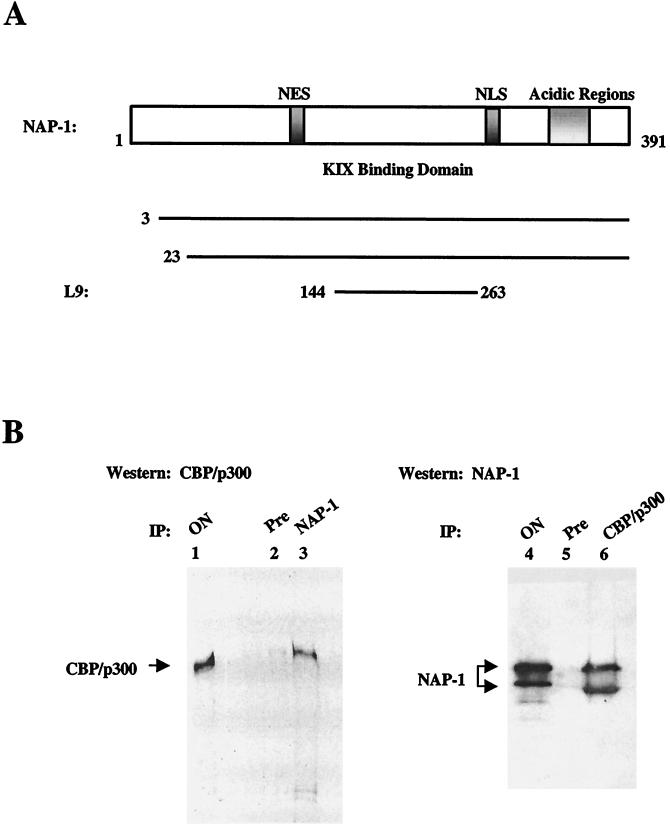
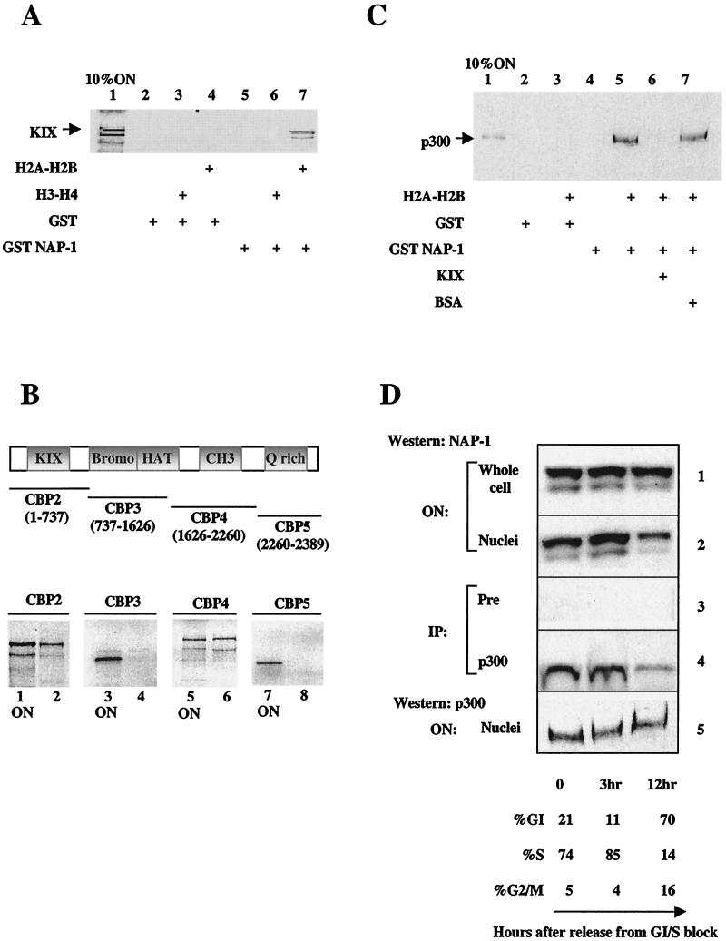
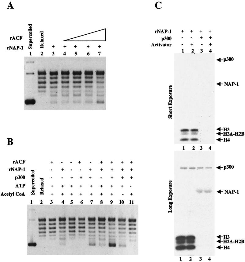
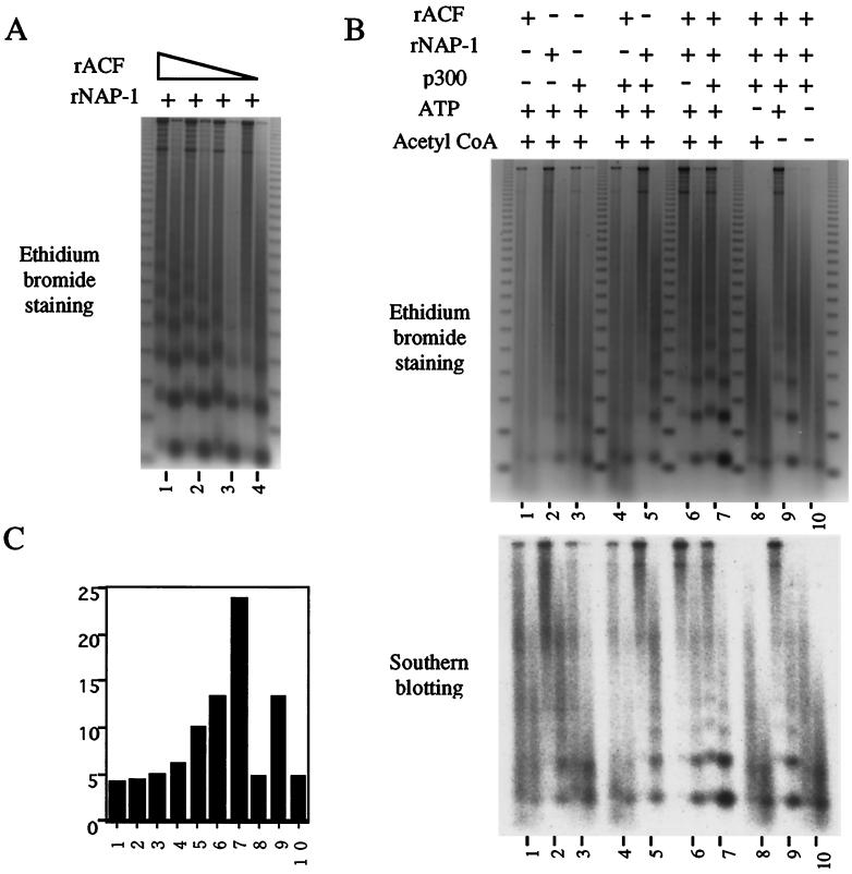
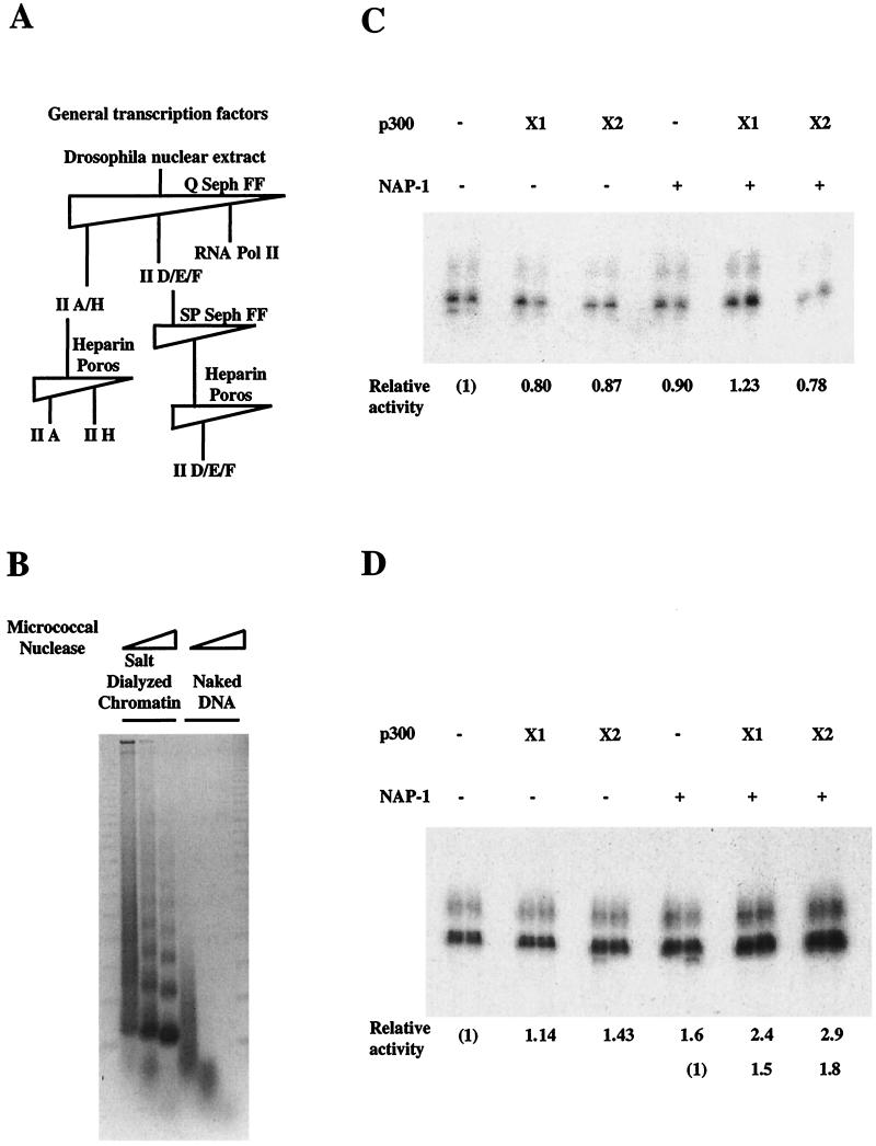
In a yeast two-hybrid screen to identify proteins that bind to the KIX domain of the coactivator p300, we obtained cDNAs encoding nucleosome assembly protein 1 (NAP-1), a 60-kDa histone H2A-H2B shuttling protein that promotes histone deposition. p300 associates preferentially with the H2A-H2B-bound form of NAP-1 rather than with the unbound form of NAP-1. Formation of NAP-1-p300 complexes was found to increase during S phase, suggesting a potential role for p300 in chromatin assembly. In micrococcal nuclease and supercoiling assays, addition of p300 promoted efficient chromatin assembly in vitro in conjunction with NAP-1 and ATP-utilizing chromatin assembly and remodeling factor; this effect was dependent in part on the intrinsic histone acetyltransferase activity of p300. Surprisingly, NAP-1 potently inhibited acetylation of core histones by p300, suggesting that efficient assembly requires acetylation of either NAP-1 or p300 itself. As p300 acted cooperatively with NAP-1 in stimulating transcription from a chromatin template in vitro, our results suggest a dual role of NAP-1-p300 complexes in promoting chromatin assembly and transcriptional activation.


Figures

FIG. 1.
FIG. 1.
NAP-1 interacts with p300 in vivo. (A) The top portion shows the structure of mammalian NAP-1 acidic regions that mediate histone H2A-H2B binding in hatched NAP-1. Consensus nuclear export (NES; aa 78 to 86) and nuclear import (NLS; aa 307 to 327) signals are indicated. The KIX binding domain (aa 144 to 263) is shown. Amino acid endpoints of independent human NAP-1 cDNA clones identified in two-hybrid screen are shown. (B) Coimmunoprecipitation studies of NAP-1 and p300 with whole-cell extracts prepared from HeLa cells. The left panel shows a Western blot assay of p300 recovered from immunoprecipitates prepared with preimmune (Pre; lane 2) or NAP-1 (lane 3) antiserum. The right panel shows a Western blot assay of NAP-1 recovered from immunoprecipitates prepared with preimmune (lane 5) or p300 (lane 6) antiserum. ON, onput protein from HeLa whole cell extract. IP, immunoprecipitate.
FIG. 2.
FIG. 2.
H2A-H2B stabilizes binding of NAP-1 to p300. (A) Pull-down assay of 35S-labeled KIX peptide (aa 553 to 679) with glutathione Sepharose beads containing GST or human GST-NAP-1. Addition of purified histone H2A-H2B or H3-H4 to binding reactions are indicated. (B) Pull-down assay of 35S-labeled CBP fragments with indicated amino acid endpoints. Fragments were incubated with GST-NAP-1 beads plus purified histone H2A-H2B. Retained fractions and 10% onput (ON) are shown. (C) GST pull-down assay of recombinant full-length p300 with resins containing GST only or GST-NAP-1 plus histone H2A-H2B are also shown. The effect of KIX polypeptide or nonspecific (bovine serum albumin; BSA) competitor (1 μg) on NAP-1-p300 complex formation is shown. ON, 10% of input recombinant p300 protein. (D) Coimmunoprecipitation studies of NAP-1 recovered from immunoprecipitates of preimmune (Pre; panel 3) or p300 antiserum (panel 4) prepared from HeLa cells synchronized by double thymidine block. The number of hours (0, 3, or 12) following release are shown below the panel. Percent of cells in G1, S, or G2 phase is estimated by fluorescence-activated cell sorter analysis of propidium iodide-stained cells. Total cellular levels of NAP-1 (panel 1), nuclear NAP-1 (panel 2), and p300 (panel 5) are shown. IP, immunoprecipitate.
FIG. 3.
FIG. 3.
p300 promotes efficient chromatin assembly in conjunction with NAP-1. (A) Chromatin assembly and supercoiling assays were performed with dNAP-1 and various concentrations of purified recombinant ACF (rACF) on a 3.2-kb plasmid template (pEcE4). (B) Using the ACF concentrations with minimal activity as described for panel A, chromatin assembly reactions were performed either in the presence or absence of indicated factors. Addition of ATP and acetyl-CoA to assembly reactions is indicated over each lane. (C) All four core histones were incubated with p300 and 3H-acetyl-CoA in the presence or absence of indicated factors. The samples were analyzed by 12% polyacrylamide gel electrophoresis, and acetylated proteins were detected by fluorography. Both short and long exposures are shown. rNAP-1, recombinant NAP-1.
FIG. 4.
FIG. 4.
p300 stimulates assembly of periodic nucleosomal arrays with NAP-1. (A) Chromatin assembly and micrococcal nuclease digestion assays were performed with NAP-1 and various concentrations of purified recombinant ACF (rACF) on an adenovirus E4 promoter template. (B and C) Chromatin assembly reactions were performed either in the presence or absence of indicated factors. Where indicated, ACF concentrations with minimal activity, as determined for panel A, were added. Addition of ATP and acetyl-CoA to assembly reactions is indicated over each lane. Two concentrations of micrococcal nuclease were employed for each condition. Digestion products were analyzed by ethidium bromide staining (top) and by Southern blot analysis (bottom), with subsequent quantitation by a PhosphorImager (C). rNAP-1, recombinant NAP-1.
FIG. 5.
FIG. 5.
NAP-1 and p300 stimulate transcription cooperatively on a chromatin template. (A) Partial purification of GTFs. Each GTF was evaluated both by Western blot and in vitro transcription assay. TFIIB was supplied as a purified recombinant protein. II A/H, TFIIA and -H; II D/E/F, TFIID, -E, and F; Seph, Sepharose. (B) Micrococcal nuclease assay of the chromatin template assembled by the salt dialysis method and naked DNA template was used for the transcription study. pEcE4 template containing five EcRE fused to the adenovirus E4 promoter was assembled into chromatin by salt dialysis. Naked DNA template (C) or salt-dialyzed chromatin (D) was incubated with various combinations of p300 and dNAP-1 as indicated in addition to the EcR/USP heterodimer, 0.7 mM acetyl-CoA, 3 mM ATP, 20-hydroxy-ecdysone ligand. After incubation chromatin template was subjected to transcription by adding partially purified GTFs and recombinant TFIIB. Fold induction over basal transcription (lane 1) was estimated by phosphorimaging. X1, one time; X2, two times.

References

    1. Adams, C. R., and R. T. Kamakaka. 1999. Chromatin assembly: biochemical identities and genetic redundancy. Curr. Opin. Genet. Dev. 9:185-190. - PubMed
    1. Akimaru, H., Y. Chen, P. Dai, D. X. Hou, M. Nonaka, S. M. Smolik, S. Armstrong, R. H. Goodman, and S. Ishii. 1997. Drosophila CBP is a coactivator of cubitus interruptus in hedgehog signalling. Nature 386:735-738. - PubMed
    1. Altman, R., and D. Kellogg. 1999. Control of mitotic events by Nap1 and the Gin4 kinase. J. Cell Biol. 138:119-130. - PMC - PubMed
    1. Arias, J., A. S. Alberts, P. Brindle, F. X. Claret, T. Smeal, M. Karin, J. Feramisco, and M. Montminy. 1994. Activation of cAMP and mitogen responsive genes relies on a common nuclear factor. Nature 370:226-228. - PubMed
    1. Asahara, H., B. Santoso, E. Guzman, K. Du, P. A. Cole, I. Davidson, and M. Montminy. 2001. Chromatin-dependent cooperativity between constitutive and inducible activation domains in CREB. Mol. Cell. Biol. 21:7892-7900. - PMC - PubMed

Publication types

MeSH terms