. 2002 May;22(9):2993-3002.
doi: 10.1128/MCB.22.9.2993-3002.2002.

Altered p27(Kip1) phosphorylation, localization, and function in human epithelial cells resistant to transforming growth factor beta-mediated G(1) arrest

Affiliations

Altered p27(Kip1) phosphorylation, localization, and function in human epithelial cells resistant to transforming growth factor beta-mediated G(1) arrest

Sandra Ciarallo et al. Mol Cell Biol. 2002 May.

Abstract

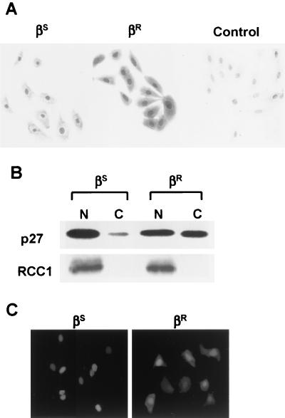
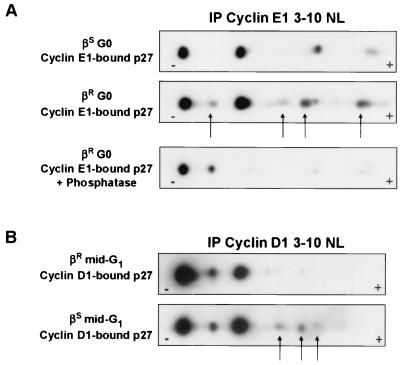
p27(Kip1) is an important effector of G(1) arrest by transforming growth factor beta (TGF-beta). Investigations in a human mammary epithelial cell (HMEC) model, including cells that are sensitive (184(S)) and resistant (184A1L5(R)) to G(1) arrest by TGF-beta, revealed aberrant p27 regulation in the resistant cells. Cyclin E1-cyclin-dependent kinase 2 (cdk2) and cyclin A-cdk2 activities were increased, and p27-associated kinase activity was detected in 184A1L5(R) cells. p27 from 184A1L5(R) cells was localized to both nucleus and cytoplasm, showed an altered profile of phosphoisoforms, and had a reduced ability to bind and inhibit cyclin E1-cdk2 in vitro when compared to p27 from the sensitive 184(S) cells. In proliferating 184A1L5(R) cells, more p27 was associated with cyclin D1-cdk4 complexes than in 184(S). While TGF-beta inhibited the formation of cyclin D1-cdk4-p27 complexes in 184(S) cells, it did not inhibit the assembly of cyclin D1-cdk4-p27 complexes in the resistant 184A1L5(R) cells. p27 phosphorylation changed during cell cycle progression, with cyclin E1-bound p27 in G(0) showing a different phosphorylation pattern from that of cyclin D1-bound p27 in mid-G(1). These data suggest a model in which TGF-beta modulates p27 phosphorylation from its cyclin D1-bound assembly phosphoform to an alternate form that binds tightly to inhibit cyclin E1-cdk2. Altered phosphorylation of p27 in the resistant 184A1L5(R) cells may favor the binding of p27 to cyclin D1-cdk4 and prevent its accumulation in cyclin E1-cdk2 in response to TGF-beta.


Figures

FIG. 1.
FIG. 1.
Cyclin-cdk2-p27 complexes and kinase activities. Cell lysates were prepared from asynchronously growing 184SS) and 184A1L5RR) cells that had similar cell cycle distributions (% S phase indicated). (A) Cyclin E1- and cyclin A-associated kinase activities. Cyclin E1 and cyclin A immunoprecipitates (IP) were assayed for associated kinase activities (left) or resolved by SDS-PAGE, transferred, and immunoblotted for cyclin and associated cdk2 (right) as described in Materials and Methods. For quantitation of the kinase reactions, radioactivity in nonspecific immune control (IgG) was subtracted and kinase activities were plotted as a percentage of the maximum (% max). Radioactivity in histone H1 bands is shown in the graph inset. (B) Cyclin E1 immunoprecipitated from asynchronously growing or TGF-β-treated 184SS) or 184A1L5RR). Complexes were resolved and proteins were detected by immunoblotting as indicated. Antibody-only control is shown on the right. (C) p27 immunoprecipitates in 184A1L5R cells contain active kinase. p27 immunoprecipitates from asynchronous HMECs with similar cell cycle distributions were assayed for cdk2 activity using histone H1 as substrate or resolved and immunoblotted for p27 and associated cdk2. Radioactivity in the nonspecific immune control (IgG) was subtracted prior to quantitation as for panel A above.
FIG. 2.
FIG. 2.
p27-cyclin D1-cdk4 assembly detected by metabolic labeling. 184S (A) and 184A1L5R (B) were grown to 60% confluence and were then arrested by EGF deprivation (G0). Cells were then transferred to complete medium and cultured for 12 h without (mid-G1) or with addition of TGF-β. At the times indicated, cells were pulse labeled with [35S]methionine and were cyclin D1 or cdk4 immunoprecipitated. Nonimmune controls (IgG) were run alongside cyclin D1-cdk4 precipitates. Cyclin D1, PCNA, cdk4, and p27 are indicated with arrows on the right. Molecular weight markers in kilodaltons (kD) are indicated on the left.
FIG. 3.
FIG. 3.
p27 binding to cyclin D1 and cyclin E1. (A) Asynchronously growing 184SS) or 184A1L5RR) cell lysates containing 200 μg of protein were serially immunodepleted of cyclin D1. Lanes 1 to 3 and 4 to 6 represent serial cyclin D1 immunoprecipitation (IP) from βS and βR cells, respectively. Immune complexes were resolved by SDS-PAGE and transferred, and blots were probed for cyclin D1 and p27. Lane C, control. (B) Cyclin D1 and p27 levels were quantitated by densitometry from different exposures of the blots in panel A, and the amounts of cyclin D1-bound p27 were corrected for the difference in total cyclin D1 between the two lines and graphed as a percentage of the maximum (% max) of that seen in the βR cells. (C) The mobilities of cyclin D1 (CycD1)- and cyclin E1 (CycE1)-bound p27 from asynchronous βS and βR cells are shown. Fifty micrograms of lysate was used for the cyclin D1 immunprecipitation, and 250 μg of lysate was used for cyclin E1 precipitation.
FIG. 4.
FIG. 4.
In vitro assays of p27's ability to bind and inhibit cyclin E1-cdk2. (A) Titrated amounts of p27 were immunoprecipitated (IP) from G0-arrested 184SS) or 184A1L5RR) cells, resolved by SDS-PAGE, and associated cellular cyclin E1 and cdk2 (endogenous) detected by immunoblotting (left panels). On the right, the indicated amounts of immunoprecipitated p27 from βS and βR cells were mixed with recombinant (recomb) cyclin E1 and GST-tagged cdk2 and were then resolved by SDS-PAGE and p27-bound endogenous (endog) and recombinant proteins detected by immunoblotting. (B) The inhibitory activity of p27 is reduced in 184A1L5R. Recombinant cyclin E1-cdk2 (recomb E/k2) was mixed with the indicated amounts of p27 immunoprecipitated from the same 184SS) or 184A1L5RR) cell lysates as for panel A above, and histone H1 kinase activity was assayed. Inhibition of E/k2 activity by the addition of p27 is shown. Radioactivity incorporated into histone H1 is shown in the autoradiograph (inset, upper right) and graphed as a percentage of the maximum (% max) uninhibited E/k2 activity. (C) The reduced affinity for E/k2 is a heat-stable property of p27 from 184A1L5R cells. The indicated amounts of immunoprecipitated p27 from βS and βR cells were boiled (100°C for 5 min). The initial amount of p27 used is indicated in the immunoblot (top left), and p27 reprecipitated after boiling is shown (lower left). The free heat-stable p27, recovered after p27 complexes were boiled, was mixed with recombinant cyclin E1 and cdk2 proteins. Cyclin E1 was then immunoprecipitated, and the amount of cyclin E1-bound cellular p27 was detected by blotting. Results shown in panels A to C are a representative of at least three experiments.
FIG. 5.
FIG. 5.
p27 shows increased cytoplasmic localization in the 184A1L5R cells. (A) Immunocytochemistry shows strong nuclear p27 staining in G0-arrested 184SS, left). p27 is localized in both nucleus and cytoplasm in G0-arrested 184A1L5R cells (βR, center). The negative control in 184A1L5R cells is shown (right). (B) p27 content of nuclear (N) and cytoplasmic (C) fractions of quiescent βS and βR cells was assayed by Western blotting. (C) βS and βR cells were transfected with fluorescence-tagged p27 (YFPp27wt), and 24 h after transfection, p27 was detected by fluorescence microscopy. Cells in panels A and C were photographed at 400× magnification.
FIG. 6.
FIG. 6.
p27 phosphorylation differs between 184S and 184A1L5R cells. p27 was immunoprecipitated (IP) using polyclonal p27 pAb5588 from G0-arrested 184SS) or 184A1L5RR) cells. For 2DIEF, complexes were resolved using the IEF strips with a linear gradient of pH 3 to 10 (3-10 L) followed by SDS-12% PAGE. Gels were transferred and p27 isoforms were detected by immunoblotting with the Transduction Laboratories p27 antibody. The IEF points (pI) of the p27 isoforms (forms 1 to 7) are indicated with arrows. In the third panel, the p27 immunprecipitates from βS and βR cells were mixed prior to 2DIEF. The bottom panel shows that, after phosphatase treatment, most of the p27 migrated at the highest IEF point, 6.5.
FIG. 7.
FIG. 7.
The phosphorylation of cyclin E1-bound p27 differs from that of cyclin D1-bound p27. The 2DIEF profile of p27 in the 184SS) or 184A1L5RR) cells was assayed in cyclin E1 immune complexes from G0 (contact inhibition for 48 h, followed by 48 h of EGF deprivation) (A) and in cyclin D1 immune complexes from mid-G1 (B). Cyclin E1-bound p27 was treated with calf intestinal alkaline phosphatase prior to 2DIEF for the lower part of panel A. Complexes were resolved on pH 3 to 10 nonlinear gradient (3-10 NL) IEF strips prior to resolution on SDS-12% PAGE and immunoblotting for p27. The arrows (A and B) indicate cyclin-associated p27 isoforms in 184A1L5R that are not detected or less abundant in 184S cells. IP, immunoprecipitation.

References

    1. Aktas, H., H. Cai, and G. M. Cooper. 1997. Ras links growth factor signaling to the cell cycle machinery via regulation of cyclin D1 and the Cdk inhibitor p27KIP1. Mol. Cell. Biol. 17:3850-3857. - PMC - PubMed
    1. Cariou, S., J. C. Donovan, W. M. Flanagan, A. Milic, N. Bhattacharya, and J. M. Slingerland. 2000. Down-regulation of p21WAF1/CIP1 or p27Kip1 abrogates antiestrogen-mediated cell cycle arrest in human breast cancer cells. Proc. Natl. Acad. Sci. USA 97:9042-9046. - PMC - PubMed
    1. Carrano, A. C., E. Eytan, A. Hershko, and M. Pagano. 1999. SKP2 is required for ubiquitin-mediated degradation of the CDK inhibitor p27. Nat. Cell Biol. 1:193-199. - PubMed
    1. Catzavelos, C., N. Bhattacharya, Y. C. Ung, J. A. Wilson, L. Roncari, C. Sandhu, P. Shaw, H. Yeger, I. Morava-Protzner, L. Kapusta, E. Franssen, K. I. Pritchard, and J. M. Slingerland. 1997. Decreased levels of the cell-cycle inhibitor p27Kip1 protein: prognostic implications in primary breast cancer. Nat. Med. 3:227-230. - PubMed
    1. Cheng, M., P. Olivier, J. A. Diehl, M. Fero, M. F. Roussel, J. M. Roberts, and C. J. Sherr. 1999. The p21(Cip1) and p27(Kip1) CDK ‘inhibitors’ are essential activators of cyclin D-dependent kinases in murine fibroblasts. EMBO J. 18:1571-1583. - PMC - PubMed

Publication types

MeSH terms

LinkOut - more resources