Case Reports
. 2002 May;70(5):1368-75.
doi: 10.1086/340390. Epub 2002 Apr 8.

Structural and functional mutations of the perlecan gene cause Schwartz-Jampel syndrome, with myotonic myopathy and chondrodysplasia

Affiliations
Case Reports

Structural and functional mutations of the perlecan gene cause Schwartz-Jampel syndrome, with myotonic myopathy and chondrodysplasia

Eri Arikawa-Hirasawa et al. Am J Hum Genet. 2002 May.

Abstract

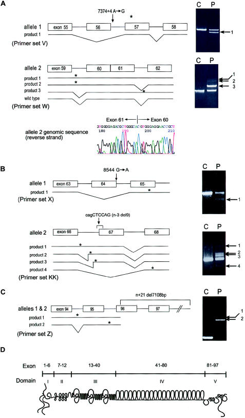
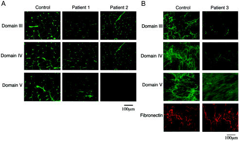
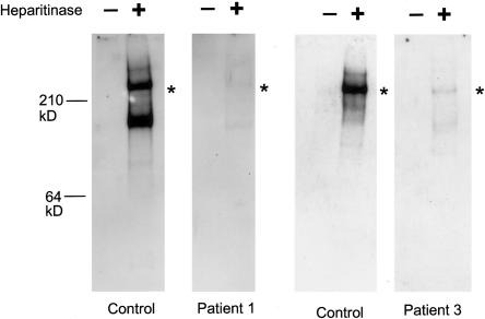
Perlecan, a large heparan sulfate proteoglycan, is a component of the basement membrane and other extracellular matrices and has been implicated in multiple biological functions. Mutations in the perlecan gene (HSPG2) cause two classes of skeletal disorders: the relatively mild Schwartz-Jampel syndrome (SJS) and severe neonatal lethal dyssegmental dysplasia, Silverman-Handmaker type (DDSH). SJS is an autosomal recessive skeletal dysplasia characterized by varying degrees of myotonia and chondrodysplasia, and patients with SJS survive. The molecular mechanism underlying the chondrodystrophic myotonia phenotype of SJS is unknown. In the present report, we identify five different mutations that resulted in various forms of perlecan in three unrelated patients with SJS. Heterozygous mutations in two patients with SJS either produced truncated perlecan that lacked domain V or significantly reduced levels of wild-type perlecan. The third patient had a homozygous 7-kb deletion that resulted in reduced amounts of nearly full-length perlecan. Unlike DDSH, the SJS mutations result in different forms of perlecan in reduced levels that are secreted to the extracellular matrix and are likely partially functional. These findings suggest that perlecan has an important role in neuromuscular function and cartilage formation, and they define the molecular basis involved in the difference in the phenotypic severity between DDSH and SJS.


Figures

Figure  1
Figure 1
Mutation analysis and schematic diagram of perlecan. A, Results of RT-PCR, which was performed with RNA from patient 1 fibroblasts, using primer sets V and W and analyzed on 1% agarose gels as described elsewhere (Arikawa-Hirasawa et al. 2001). Sequencing revealed that product 1 from allele 1 was missing the entire exon 56 sequence and that products 1–3 from allele 2 retained intron sequences. Product 3 of allele 2 contains the sequence of intron 61, whereas products 1 and 2 contain either both introns 59 and 61 or intron 59, respectively. Exon 56 skipping in allele 1 causes frameshift and is predicted to produce a premature termination codon in exon 57. The intron 59 retention of products 1 and 2 from allele 2 is predicted to produce a premature termination codon within intron 59, and the intron 61 retention of product 3 is predicted to produce a termination codon within intron 61. Genomic sequencing identified heterozygous mutations in an A→G transition at the +4 position (donor site) of intron 56 (7374+4 A→G) in allele 1. Allele 2 showed a complete loss of intron 60, resulting in fusion of exons 60 and 61 in the perlecan gene. The exon-fusion mutation created aberrant transcripts, products 1–3, but is also predicted to produce the wild-type product. Since primer set W does not distinguish the wild-type product derived from alleles 1 or 2, we used a primer set from exons 56 and 62 to exclude a wild-type transcript from allele 1, in which exon 56 is missing and confirmed the presence of the wild-type transcript together with aberrant splicing transcripts produced by allele 2 (data not shown). B, Patient 2. RT-PCR was performed with RNA from skeletal muscle tissues of patient 2, using primer sets X and KK. Sequencing of product 1 with primers X revealed exon 64 skipping, and products 1, 2, and 3 with primers KK contained total or partial retention of intron 66. Product 4 with primers KK showed exon 67 skipping. Genomic sequencing revealed a heterozygous mutation in a G→A transition at the donor site of exon 64 (8564 G→A) in allele 1 and a 9-bp deletion (cagCTCCAG) at the acceptor junction of intron 66 and exon 67 in allele 2 (n−3 del9). C, Patient 3. RT-PCR was performed with RNA from patient 3 fibroblasts using primer set Z. Product 1 contained sequences including introns 94 and 95. Product 2, a major product, contained intron 95. Sequencing of the genomic PCR product revealed a 7,108-bp deletion beginning at the 5′ portion of exon 96 and extending to the 3′ flanking sequence of HSPG2. The deletion results in aberrant splicing, including exon skipping and intron retention. Asterisk (*) indicates a premature termination codon. Arrows indicate abnormally sized RT-PCR products. D,Schematic diagram of perlecan and its exon and domain structure. Primer sequences for RT-PCR are as follows: V (forward, ATCACGGTCACAGTAACTGGGACC; reverse, CCTGCACCGTTACTGACGTG); KK (forward, ATGGCACAAGCGTGGAGGAAACC; reverse, AGGCTTCTTGCTCAGGGCCTGG); W (forward, ATCCAGCAGCGCCTTAGTGG; reverse, CATGCCCATCAGAATTGAGTCAT); X (forward, CTCAACAACATCG ATGCCCTGGAG; reverse, CTCCAGCCCAGGACCCATTCCT); and Z (forward, AGGCAAGGACTTCATCAGCCTCGGG; reverse, TCGACTTGGATGGAACCTCTGCGG).
Figure  2
Figure 2
Immunostaining of perlecan in muscle tissues and cultured fibroblasts. A, Muscle tissue from patients 1 and 2 and from an unaffected control subject, stained with domain-specific anti-perlecan antibodies as described elsewhere (Arikawa-Hirasawa et al. 2001, 2002). In patient 1, antibodies to domains III–V stained the basal lamina of the muscle, whereas, in patient 2, domain V staining was absent. The staining in muscle tissue of patient 1 is significantly reduced. B, Cultured fibroblasts from patient 3 stained with domain-specific anti-perlecan antibodies and with anti-fibronectin polyclonal antibodies. Domains III–V stained the extracellular matrix at significantly reduced levels compared to control fibroblasts, whereas fibronectin stained strongly in both.
Figure  3
Figure 3
Perlecan secreted by cultured fibroblasts in patients 1 and 3. Media from cultured normal or patient fibroblasts were concentrated, were then adjusted to contain 0.35g CsCl/g of 4M guanidine, and were centrifuged for 72 h at 40,000 RPM in a 50-Ti rotor at 12°C. A proteoglycan-containing fraction was pooled, concentrated, and digested with 0.1 mU heparitinase and 0.25 mU chondroitinase ABC (Seikagaku). Digested and undigested samples were electrophoresed in 3%–8% polyacrylamide tris-acetate gels, were transferred to nitrocellulose, and were blotted with polyclonal antibodies to the perlecan protein core. In patient 1, a perlecan protein core of ∼400 kD was detected after heparitinase digestion, but the amount was reduced significantly, correlating with the reduced amount of the wild-type perlecan transcript. Heparitinase treatment is necessary for the blotting, because perlecan-containing heparan sulfate chains do not transfer well onto a membrane because of the negatively charged heparan sulfate chains. As with patient 1, patient 3 fibroblasts secreted the 400-kD perlecan core protein in much more reduced amounts than did the control cells. The truncated perlecan in patient 3 is missing only ∼35–64 amino acids of the most C-terminal part of domain V, suggesting the mutant perlecan to be almost the same size as the wild-type perlecan. Asterisks (*) indicate positions of the ∼400-kD protein core.

References

Electronic-Database Information

    1. GenBank, http://www.ncbi.nlm.nih.gov/GenBank/ (for the sequence of HSPG2 cDNA [accession number M852890] and for HSPG2-containing human chromosome 1 working draft sequence segment [accession number NT_004576])
    1. Online Mendelian Inheritance in Man (OMIM), http://www.ncbi..nih.gov/Omin/ (for SJS [MIM 255800] and DDSH [MIM 224410])

References

    1. Aberfeld DC, Hinterbuchner LP, Schneider M (1965) Myotonia, dwarfism, diffuse bone disease and unusual ocular and facial abnormalities (a new syndrome). Brain 88:313–322 - PubMed
    1. Aberfeld DC, Namba T, Vye MV, Grob D (1970) Chondrodystrophic myotonia: report of two cases—myotonia, dwarfism, diffuse bone disease, and unusual ocular and facial abnormalities. Arch Neurol 22:455–462 - PubMed
    1. Arikawa-Hirasawa E, Rossi SG, Rotundo RL, Yamada Y (2002) Absence of acetylcholinesterase at the neuromuscular junctions of perlecan-null mice. Nat Neurosci 5:119–123 - PubMed
    1. Arikawa-Hirasawa E, Watanabe H, Takami H, Hassell JR, Yamada Y (1999) Perlecan is essential for cartilage and cephalic development. Nat Genet 23:354–358 - PubMed
    1. Arikawa-Hirasawa E, Wilcox WR, Le AH, Silverman N, Govindraj P, Hassell JR, Yamada Y (2001) Dyssegmental dysplasia, Silverman-Handmaker type, is caused by functional null mutations of the perlecan gene. Nat Genet 27:431–434 - PubMed

Publication types

MeSH terms

Associated data

LinkOut - more resources