. 2002 Apr;160(4):1425-33.
doi: 10.1016/S0002-9440(10)62568-0.

Evidence for an active inflammatory process in the hibernating human myocardium

Affiliations

Evidence for an active inflammatory process in the hibernating human myocardium

Nikolaos G Frangogiannis et al. Am J Pathol. 2002 Apr.

Abstract

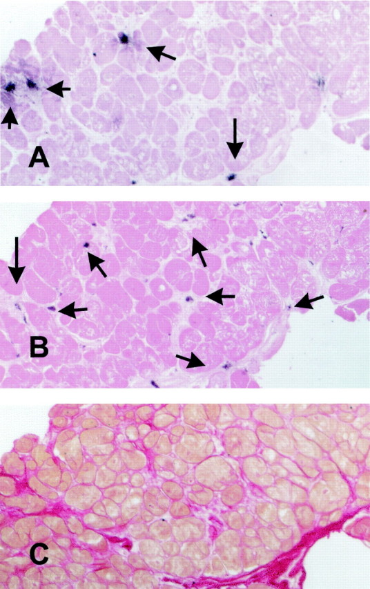
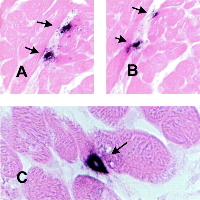
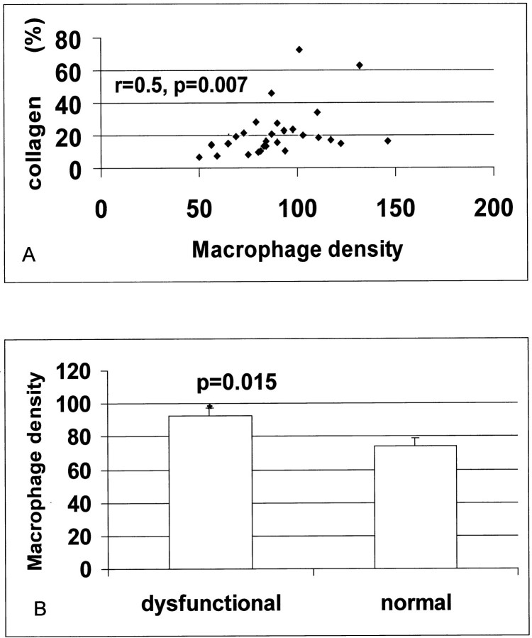
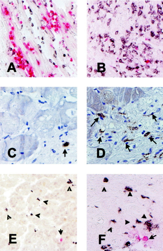
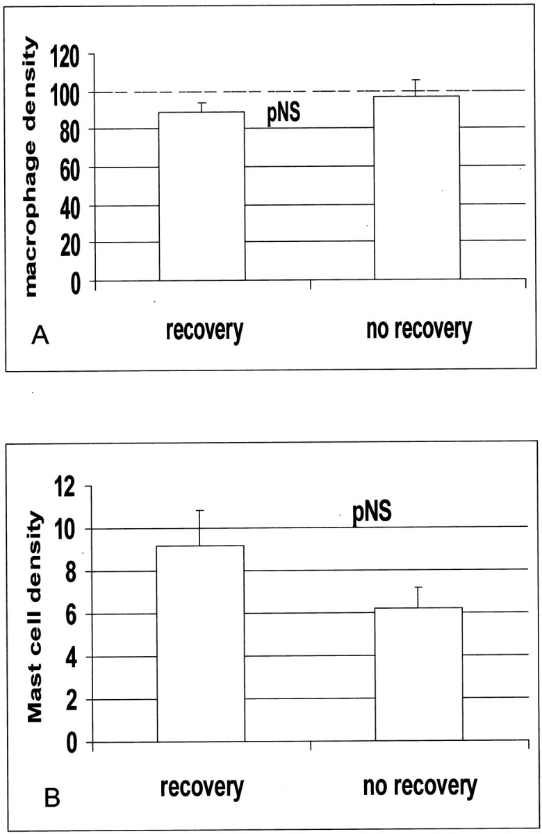
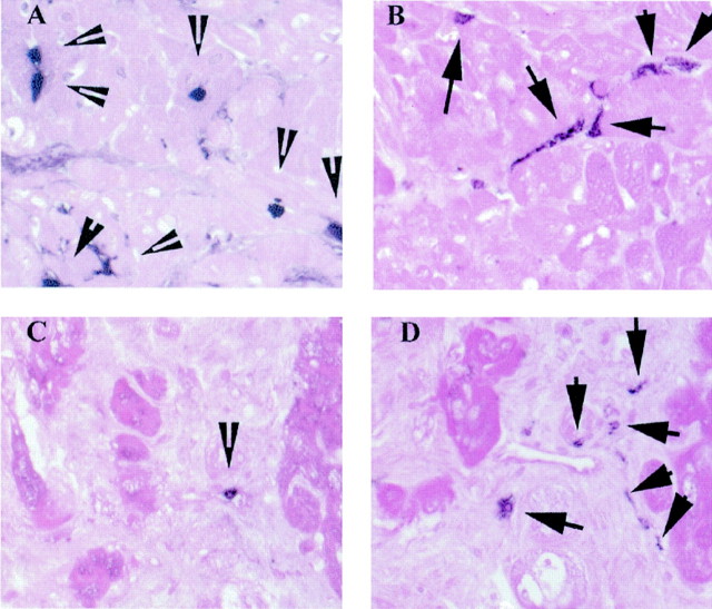
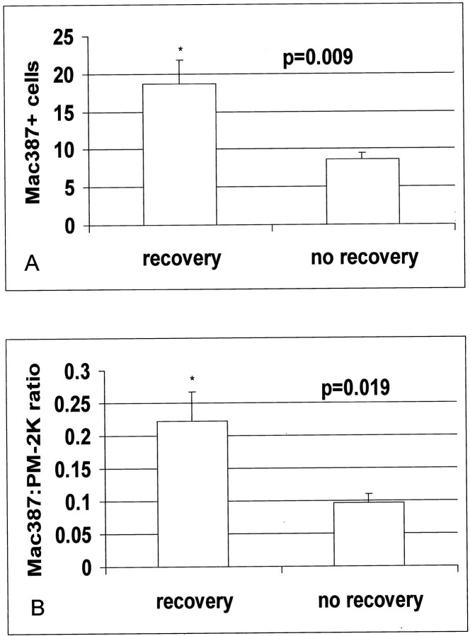
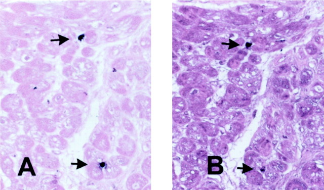
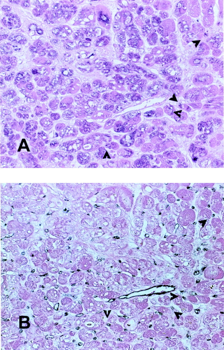
Myocardial hibernation refers to a state of prolonged impairment of left ventricular function in the presence of coronary artery disease, which may be reversed by revascularization. In this study we present evidence for a local inflammatory reaction in hibernating myocardial segments from patients undergoing coronary revascularization. We obtained transmural myocardial biopsies guided by transesophageal echocardiography from patients with ischemic ventricular dysfunction undergoing bypass surgery. Among the 28 biopsied segments included in the study, 23 showed evidence of systolic dysfunction. The majority of dysfunctional segments (85.7%) were viable ((201)Tl uptake >/= 60%). The samples were stained with markers for mast cells, mature resident macrophages, and the monoclonal antibody Mac387 that labels newly recruited myeloid cells. Dysfunctional segments showed more extensive fibrosis and higher macrophage density than normal segments. Among the 23 dysfunctional segments, 12 recovered function as assessed with echocardiograms 3 months after revascularization. Segments with postoperative functional recovery had comparable macrophage and mast cell density with those showing persistent dysfunction. However, biopsied segments that subsequently recovered function contained significantly higher numbers of newly recruited Mac387-positive leukocytes (18.7 +/- 3.1 cells/mm(2), n = 12 versus 8.6 +/- 0.9 cells/mm(2), n = 11; P = 0.009). In addition, monocyte chemotactic protein-1, a potent mononuclear cell chemoattractant, was predominantly expressed in segments with recovery of function. Myocardial hibernation is associated with an inflammatory response leading to active leukocyte recruitment. Dysfunctional myocardial segments that show an active inflammatory reaction have a greater potential for recovery of function after revascularization. We postulate that revascularization may promote resolution of the ongoing inflammation, preventing further tissue injury and fibrosis.


Figures

Figure 1.
Figure 1.
Labeling of mast cells and macrophages in the human myocardium. Serial myocardial sections were stained for tryptase (A) to identify mast cells (arrows) and for the monoclonal antibody PM-2K (B) to label mature resident macrophages (arrows). Both slides were counterstained with eosin. Adjacent sections were stained with picrosirius red for collagen (C). Original magnifications, ×200.
Figure 2.
Figure 2.
Mast cells in the human heart are identified as granular interstitial cells with intense immunoreactivity for tryptase (A and C) and chymase (B).
Figure 3.
Figure 3.
Macrophage density correlates with collagen deposition and is higher in dysfunctional myocardial segments. A significant correlation between macrophage density and collagen percent staining was noted (r = 0.5, P = 0.007) (A). Macrophage numbers were higher in dysfunctional myocardial segments when compared with segments showing normal regional function (*, P = 0.015) (B).
Figure 4.
Figure 4.
Use of the monoclonal antibody Mac387 as a marker of acute inflammation in the heart. A: Canine myocardial infarction after 1 hour of circumflex coronary artery occlusion and 24 hours of reperfusion. Peroxidase-based immunohistochemistry for the macrophage-specific antibody PM-2K (black) was followed by alkaline phosphatase-based staining for Mac387 a marker of newly recruited myeloid cells (red). Note the abundance of Mac387-positive cells in the infarcted area. B: Canine infarct after 1 hour of coronary occlusion and 7 days of reperfusion. The healing infarct is now filled with mature macrophages (black); whereas only a few newly recruited leukocytes are noted (red). C: Immunohistochemical staining of a biopsied human myocardial sample with the Mac387 antibody followed by hematoxylin counterstaining identified newly recruited leukocytes. D: Immunostaining of a biopsied human myocardial sample with the macrophage-specific antibody PM-2K labeled mature interstitial macrophages. E and F: Dual immunohistochemical staining combining peroxidase-based staining for PM-2K (black) and alkaline phosphatase-based staining for Mac387 (red) demonstrating that macrophages (arrowheads) significantly outnumbered newly recruited leukocytes (arrow). Original magnifications: ×200 (A, B, E); ×400 (C, D, F).
Figure 5.
Figure 5.
Macrophage (A) and mast cell (B) density is similar in dysfunctional segments with and without recovery of function after revascularization.
Figure 6.
Figure 6.
Immunohistochemical labeling of newly recruited Mac387-positive myeloid cells (A and C) and mature PM-2K-positive resident macrophages (B and D) in the myocardium. Representative sections from a dysfunctional segment that showed recovery of function after revascularization are shown (A and B). Note the significant number of newly recruited Mac-387-positive cells (A, arrowheads), compared to the number of mature resident macrophages (B, arrows). In contrast, segments with persistent dysfunction (C and D) had rare newly recruited cells (C, arrowhead) and a high number of resident macrophages (D, arrows). The slides were counterstained with eosin. Original magnifications, ×400.
Figure 7.
Figure 7.
A: Segments with recovery of function show higher numbers of Mac387-positive cells when compared with those without recovery (P = 0.009). B: In addition, the ratio of newly recruited cells to mature macrophages was significantly higher in segments with recovery (P = 0.019).
Figure 8.
Figure 8.
Newly recruited leukocytes express MMP-9, exhibiting a protein-degrading migratory phenotype. Immunohistochemical staining of a biopsied myocardial sample with Mac387 detects two newly recruited leukocytes (arrows) (A). Serial section staining demonstrates MMP-9 expression by these cells (arrows) (B). Original magnifications, ×400.
Figure 9.
Figure 9.
Immunohistochemical staining for MCP-1 in a myocardial segment with recovery of function after revascularization (A). Diffuse staining is noted in many cardiomyocytes and occasional microvascular endothelial cells (arrows) identified by serial section staining with a monoclonal antibody to CD31 (B). Original magnifications, ×400.

References

    1. Rahimtoola SH: Concept and evaluation of hibernating myocardium. Annu Rev Med 1999, 50:75-86 - PubMed
    1. Rahimtoola SH: A perspective on the three large multicenter randomized clinical trials of coronary bypass surgery for chronic stable angina. Circulation 1985, 72:V123-V135 - PubMed
    1. Heusch G: Hibernating myocardium. Physiol Rev 1998, 78:1055-1085 - PubMed
    1. Vanoverschelde JL, Pasquet A, Gerber B, Melin JA: Pathophysiology of myocardial hibernation. Implications for the use of dobutamine echocardiography to identify myocardial viability. Heart 1999, 82(Suppl 3):III1-III7 - PMC - PubMed
    1. Elsasser A, Schaper J: Hibernating myocardium: adaptation or degeneration? Basic Res Cardiol 1995, 90:47-48 - PubMed

Publication types