. 2002 Apr 15;22(8):3161-73.
doi: 10.1523/JNEUROSCI.22-08-03161.2002.

Dividing precursor cells of the embryonic cortical ventricular zone have morphological and molecular characteristics of radial glia

Affiliations

Dividing precursor cells of the embryonic cortical ventricular zone have morphological and molecular characteristics of radial glia

Stephen C Noctor et al. J Neurosci. .

Abstract

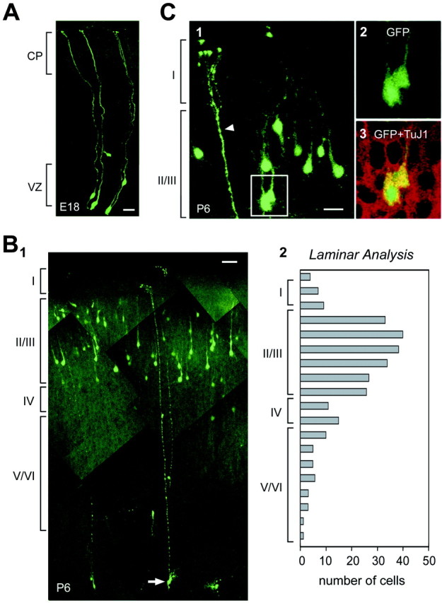
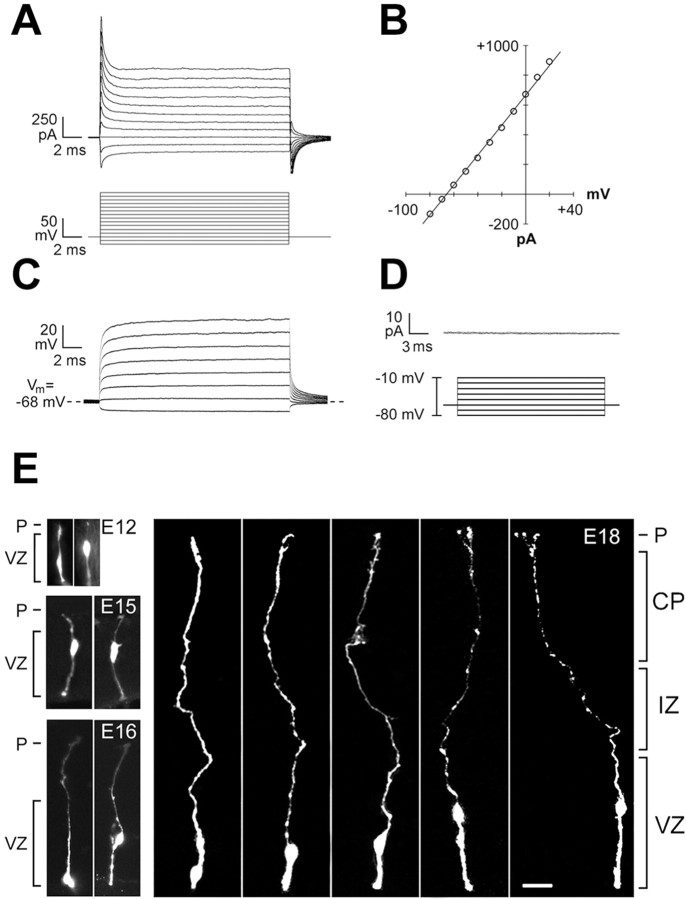
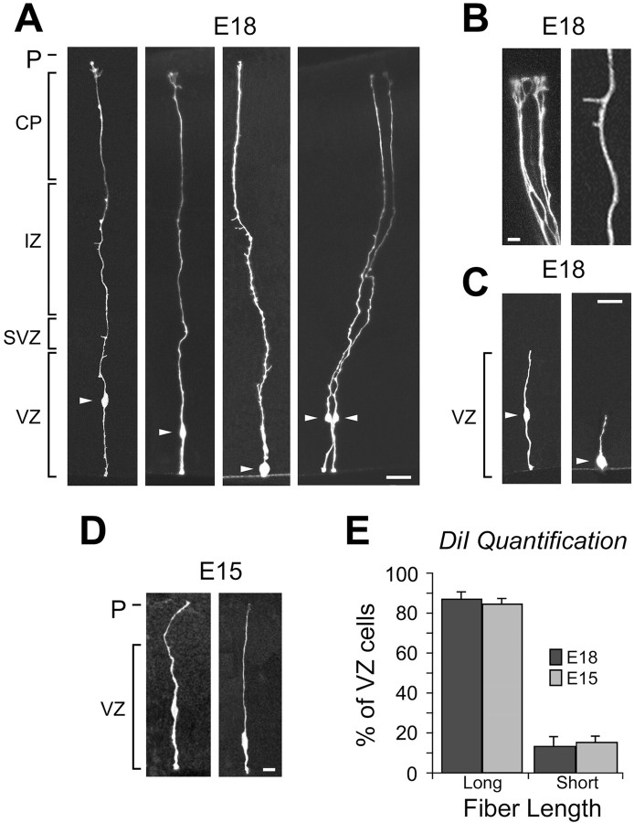
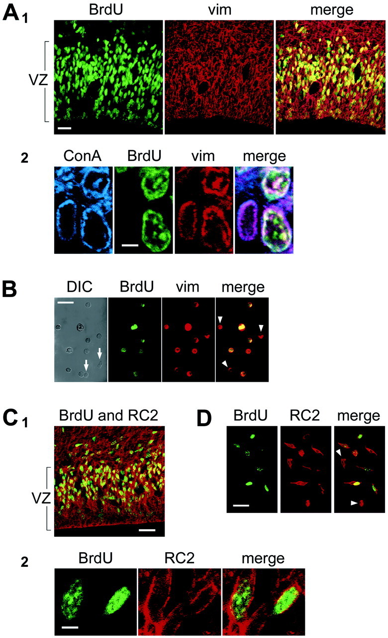
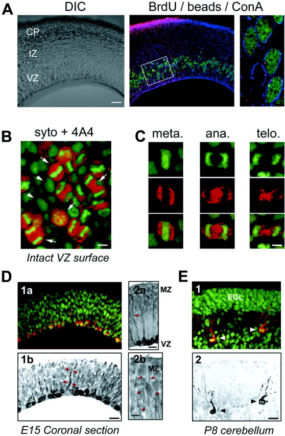
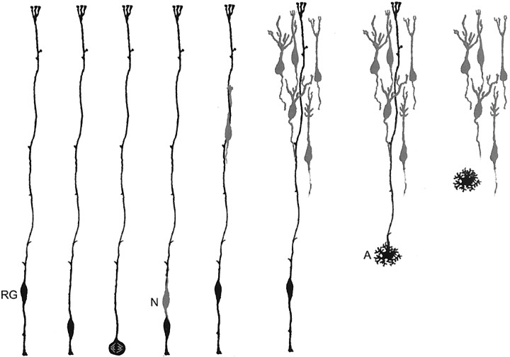
The embryonic ventricular zone (VZ) of the cerebral cortex contains migrating neurons, radial glial cells, and a large population of cycling progenitor cells that generate newborn neurons. The latter two cell classes have been assumed for some time to be distinct in both function and anatomy, but the cellular anatomy of the progenitor cell type has remained poorly defined. Several recent reports have raised doubts about the distinction between radial glial and precursor cells by demonstrating that radial glial cells are themselves neuronal progenitor cells (Malatesta et al., 2000; Hartfuss et al., 2001; Miyata et al., 2001; Noctor et al., 2001). This discovery raises the possibility that radial glia and the population of VZ progenitor cells may be one anatomical and functional cell class. Such a hypothesis predicts that throughout neurogenesis almost all mitotically active VZ cells and a substantial percentage of VZ cells overall are radial glia. We have therefore used various anatomical, immunohistochemical, and electrophysiological techniques to test these predictions. Our data demonstrate that the majority of VZ cells, and nearly all mitotically active VZ cells during neurogenesis, both have radial glial morphology and express radial glial markers. In addition, intracellular dye filling of electrophysiologically characterized progenitor cells in the VZ demonstrates that these cells have the morphology of radial glia. Because the vast majority cycling cells in the cortical VZ have characteristics of radial glia, the radial glial precursor cell may be responsible for both the production of newborn neurons and the guidance of daughter neurons to their destinations in the developing cortex.


Figures

Fig. 1.
Fig. 1.
GFP-retroviral labeling at E17 demonstrates radial glia at 30 hr and upper layer cortical neurons at P6. A, Representative labeling of radial glia 30 hr after infection.B1, At P6, GFP-expressing neurons are distributed mainly in the upper layers of cortex.B2, Histogram of the laminar distribution of labeled cells confirms localization to upper cortical layers. C1, At P6, many pyramidal-like cortical cells are GFP positive, as are the apical processes of transforming radial glia (arrowhead).C3, Optical sections through the GFP-expressing cortical cells shown inC2 demonstrate the presence of the neuronal marker TuJ1. Layers I–VI are indicated. Scale bars:A, 10 μm; B1, 20 μm; C, 20 μm.
Fig. 2.
Fig. 2.
All recorded cells had the expected physiological properties of precursor cells. A, Typical recordings of current responses to a series of voltage steps in voltage-clamp mode demonstrate low input resistance. B, Current–voltage is linear between −90 mV and +10 mV in the same cell as A.C, Current responses to a series of voltage steps recorded in current-clamp mode. D, Leak-subtracted voltage-clamp recordings confirm the absence of voltage-gated sodium conductances. E, Representative cells recorded and filled at E12, E15, E16, and E18. Five representative recorded cells shown atE18 exhibit radial glial morphology, with an endfoot on the ventricle, a cell body within the VZ, and a radial process extending to the pia (P). Scale bar, 10 μm.
Fig. 3.
Fig. 3.
Random labeling of VZ cells by DiOlistics suggests that most of the VZ cells are radial glia. A, Shown are labeled cells at E18 with somata (arrowheads) in the upper, middle, and lower zones of the VZ and long fibers extending to the pia. B, Cells identified by DiOlistic labeling have expected morphological features of radial glial cells, such as branched pial endfeet of the long radial fiber (left) and small side branches (right). C, A small percentage of cells with processes restricted to the VZ are also observed, with cell bodies (arrowheads) in the middle (left) and lower (right) zones of the VZ. D, The majority of VZ cells labeled by DiOlistics at E15 resembled these two examples with long radial fibers. E, Quantitative analysis of the percentage of cells with long pially directed fibers (Long) versus cells with fibers restricted to the VZ (Short) identified by random DiOlistic labeling of the VZ surface at E15 and E18. Scale bars: A,C, 10 μm; B, D, 5 μm.CP, Cortical plate; IZ, intermediate zone; P, pia; SVZ, subventricular zone.
Fig. 4.
Fig. 4.
S-phase cells in the VZ express the radial glial markers vimentin and RC2.A1, BrdU labeling (BrdU, green) in E18 rat tissue fixed 1 hr after BrdU injection to identify S-phase cells. Vimentin labeling (vim, red) labels most cells in the VZ. Overlay of the two channels is shown in the right panel(merge). A2, Colocalization of BrdU and vimentin labeling in brain slices was confirmed using a third marker, concanavalin A (ConA,blue) to label cellular membranes. Overlay of the BrdU, ConA, and vimentin channels is shown on the right(merge). B, BrdU-positive dissociated single cells are also vimentin positive. Some cells (arrows in DIC panel) do not express either marker, and some vimentin-positive cells do not express BrdU (arrowheads). C1, In E15 mouse brain slices, BrdU (green) colocalizes with the murine radial glial marker, RC2 (red). C2, Colocalization of BrdU and RC2 was determined at higher magnification than in C1, as shown here.D, BrdU-positive dissociated single cells are also RC2 positive. Scale bars: A, C, 15 μm;B, 5 μm; D, 20 μm.
Fig. 5.
Fig. 5.
M-phase cells in the VZ express the radial glial marker 4A4, and cells in S-phase and M-phase have radial glial morphology. A, BrdU-positive cells (green) are labeled by fluorescent microspheres (beads, red) placed on the pia. Colabeling (shown at E17) is confirmed by ConA staining of cell borders (blue). Cells were analyzed in the VZ underlying pial bead application sites (box). Most BrdU-positive cells contain beads as shown in the right panel.B, Confocal sections of the ventricular surface from intact cortical slabs (shown at E15) demonstrate localization of 4A4 immunostaining (red) in cells identified as M-phase cells by the pattern of Syto-11 labeling (green).Arrows identify cells in different stages of (Figure legend continued.) M-phase that are 4A4 positive. Arrowheads identify cells without condensed chromatin that are not in M-phase and do not express 4A4. C, Cells in specific stages of M-phase, including metaphase, anaphase, and telophase, are clearly identified by Syto-11 chromatin morphology (green) and have surrounding 4A4 fluorescence (red). D1a, Overlay of Syto-11 (green) and 4A4 (red) fluorescence in a coronal section at E15.D1b, 4A4 staining in this same section (black) shows multiple cells labeled at the ventricular border and 4A4-positive radial fibers extending toward the pia (arrowheads). D2a, Radial processes of 4A4-positive cells (arrowhead) extend to the pia and marginal zone (MZ). D2b, Faint 4A4 labeling is present in distal fibers that reach the MZ (arrowheads). E1, Overlay of Syto-11 (green) and 4A4 (red) in P8 cerebellum demonstrates colabeling in Bergmann radial glia (arrowheads). Granule cell precursors in the external granule layer (EGL) do not express 4A4.E2, 4A4-positive staining (black) in the same section as E1 demonstrates morphology of M-phase Bergmann radial glia (arrowheads). Scale bars: A, 100 μm; B, 5 μm;C, 4 μm; D1,D2a, E, 10 μm;D2b, 5 μm.
Fig. 6.
Fig. 6.
Schematic hypothesis for neuronal generation in the neocortex. Our model proposes that one population, the radial glial cell (RG), both generates neurons and guides neuronal migration. Newborn neurons (N) generated by radial glial precursor cells ascend along the radial fiber of the parental radial glial cell. A series of asymmetric radial glial cell divisions produces a clone of cortical pyramidal neurons. After neurogenesis, some radial glial cells transform into astrocytes (A).

References

    1. Altman J, Bayer SA. Vertical compartmentation and cellular transformations in the germinal matrices of the embryonic rat cerebral cortex. Exp Neurol. 1990;107:23–35. - PubMed
    1. Alvarez-Buylla A, Buskirk DR, Nottebohm F. Monoclonal antibody reveals radial glia in adult avian brain. J Comp Neurol. 1987;264:159–170. - PubMed
    1. Alvarez-Buylla A, Theelen M, Nottebohm F. Proliferation “hot spots” in adult avian ventricular zone reveal radial cell division. Neuron. 1990;5:101–109. - PubMed
    1. Alvarez-Buylla A, Garcia-Verdugo JM, Tramontin AD. A unified hypothesis on the lineage of neural stem cells. Nat Rev Neurosci. 2001;2:287–293. - PubMed
    1. Anderson S, Mione M, Yun K, Rubenstein JL. Differential origins of neocortical projection and local circuit neurons: role of Dlx genes in neocortical interneuronogenesis. Cereb Cortex. 1999;9:646–654. - PubMed

Publication types

MeSH terms