. 2002 Apr 15;22(8):3234-43.
doi: 10.1523/JNEUROSCI.22-08-03234.2002.

Cholinergic changes in the APP23 transgenic mouse model of cerebral amyloidosis

Affiliations

Cholinergic changes in the APP23 transgenic mouse model of cerebral amyloidosis

Sonia Boncristiano et al. J Neurosci. .

Abstract

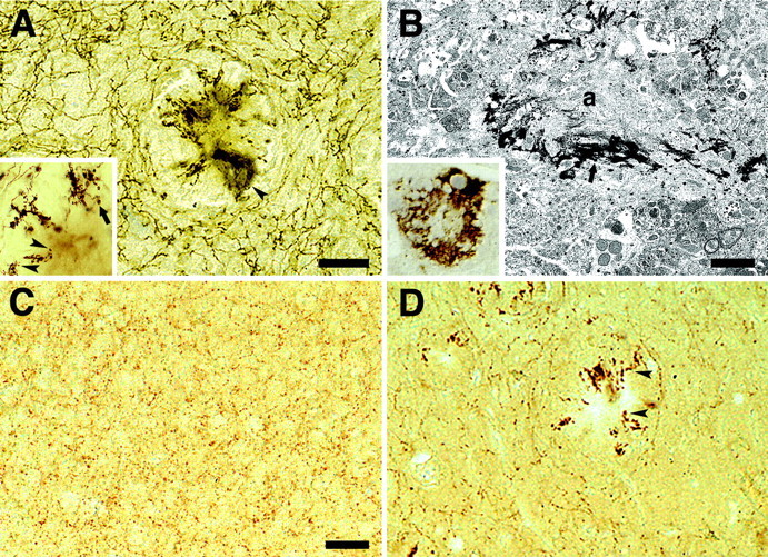
Alzheimer's Disease (AD) is a neurodegenerative disorder that is characterized by extracellular deposits of amyloid-beta peptide (Abeta) and a severe depletion of the cholinergic system, although the relationship between these two events is poorly understood. In the neocortex, there is a loss of cholinergic fibers and receptors and a decrease of both choline acetyltransferase (ChAT) and acetylcholinesterase enzyme activities. The nucleus basalis of Meynert (NBM), which provides the major cholinergic input to the neocortex, undergoes profound neuron loss in AD. In the present study, we have examined the cholinergic alterations in amyloid precursor protein transgenic mice (APP23), a mouse model of cerebral beta-amyloidosis. In aged APP23 mice, our results reveal modest decreases in cortical cholinergic enzyme activity compared with age-matched wild-type mice. Total cholinergic fiber length was more severely affected, with 29 and 35% decreases in the neocortex of aged APP23 mice compared with age-matched wild-type mice and young transgenic mice, respectively. However, there was no loss of cholinergic basal forebrain neurons in these aged APP23 mice, suggesting that the cortical cholinergic deficit in APP23 mice is locally induced by the deposition of amyloid and is not caused by a loss of cholinergic basal forebrain neurons. To study the impact of cholinergic basal forebrain degeneration on cortical amyloid deposition, we performed unilateral NBM lesions in adult APP23 mice. Three to 8 months after lesioning, a 38% reduction in ChAT activity and significant cholinergic fiber loss were observed in the ipsilateral frontal cortex. There was a 19% decrease in Abeta levels of the ipsilateral compared with contralateral frontal cortex with no change in the ratio of Abeta40 to Abeta42. We conclude that the severe cholinergic deficit in AD is caused by both the loss of cholinergic basal forebrain neurons and locally by cerebral amyloidosis in the neocortex. Moreover, our results suggest that disruption of the basal cholinergic forebrain system does not promote cerebral amyloidosis in APP23 transgenic mice.


Figures

Fig. 1.
Fig. 1.
Amyloid deposits and cholinergic disruption in neocortex of aged APP23 mice. A, No amyloid is detected in 24-month-old wild-type mice. B, Immunostaining for Aβ reveals numerous compact amyloid plaques, diffuse amyloid (arrowheads), and amyloid deposits in vessels (arrow) in neocortex of a 24-month-old APP23 mouse.C, D, Histochemical staining for AChE reveals a disruption and a decrease in cholinergic fiber density in neocortex of 24-month-old APP23 mice (D) compared with 24 month-old control mice (C). The disruption is most evident around plaques but occurs throughout the neocortex. Scale bars, 120 μm.
Fig. 2.
Fig. 2.
Cholinergic disruption and dystrophy in APP23 neocortex. A, AChE-positive fibers around an amyloid plaque with diffuse staining of the amyloid cores in the neocortex of an aged APP23 mouse (arrowhead).Inset, High-power analysis of the abnormal and often swollen AChE-positive fibers with terminal large boutons in vicinity of the amyloid plaque (arrowheads). Fibers frequently grow toward the amyloid but then form loops or sharply turn around to grow away from the amyloid to turn later again toward the amyloid (arrow). B, High-power analysis of diffuse AChE-staining associated with the amyloid cores.Inset, AChE staining in semithin sections suggests an association of AChE with the amyloid core. Subsequent ultrastructural analysis confirms that AChE reactivity (arrow) is associated with amyloid fibers (a).C, Immunostaining for ChAT reveals dense punctate staining of cholinergic boutons in the frontal cortex of an aged wild-type mouse. D, In contrast, a loss of cholinergic boutons is apparent in aged APP23 mice. Dystrophic ChAT-positive boutons and neuritic structures are present around amyloid plaques (arrowheads). Scale bars: A, 50 μm;B, 1.6 μm; C, D, 50 μm.
Fig. 3.
Fig. 3.
AChE-positive fiber length and amyloid load in neocortex of APP23 mice. A, Total length of AChE-positive fibers in neocortex of young (6 months), adult (15 months), and aged (24 months) APP23 mice (black bars) and age-matched wild-type mice (open bars). Aged transgenic mice had a significant loss of fiber length compared with aged wild-type mice (**p < 0.01) and with young transgenic mice (**p < 0.01). Indicated is the mean ± SEM for one hemisphere only. B, A significant negative correlation between AChE-positive fiber length and amyloid load was apparent when adult (circles) and aged (squares) APP23 mice were combined.
Fig. 4.
Fig. 4.
ChAT-positive neurons in the basal forebrain of APP23 mice. A, B, No apparent difference in neuron number was observed in the NBM of 27-month-old APP23 mice (A) compared with aged-matched wild-type mice (B). C, D, Similarly, no apparent difference in ChAT-positive neuron number in the MS was noted between 27-month-old APP23 (C) mice and age-matched control mice (D). For quantification, see Figure5. Scale bar, 50 μm.
Fig. 5.
Fig. 5.
Number and volume of cholinergic neurons in the basal forebrain of APP23 mice. Number (A) and volume (C) of ChAT-positive neurons in the NBM complex in APP23 mice (black bars) and wild-type mice (open bars). No significant difference between transgenic and wild-type mice was noted. A similar analysis was done for the number (B) and volume (D) of ChAT-positive neurons in the MS–VDB in APP23 mice (black bars) and wild-type mice (open bars). For neuron volume, a significant reduction of 38 and 42% was found in the 8-month-old and 27-month-old APP23 mice, respectively (*p < 0.05). Data are mean ± SEM. Indicated values are for one hemisphere only.
Fig. 6.
Fig. 6.
Amyloid load in the frontal cortex after NBM lesions. A, B, Double labeling for AChE (brown) and Aβ (blue) in a 13-month-old APP23 mouse 3 months after a unilateral NBM lesion reveals a considerable loss of cholinergic fibers in the ipsilateral (B) compared with contralateral (A) frontal cortex. Stereological assessment revealed a 22% reduction of amyloid deposition in the ipsilateral compared with contralateral side. Scale bar: A,B, 90 μm. C, Western blotting of cortex homogenates. Samples were run in pairs, i.e., contralateral side (c) versus ipsilateral side (i). Shown are three mice. Results for all the mice revealed a 19% decrease of Aβ40 between the two sides. No difference in the ratio between Aβ40 and Aβ42 was found between contralateral and ipsilateral side.

References

    1. Andra K, Abramowski D, Duke M, Probst A, Wiederhold KH, Burki K, Goedert M, Sommer B, Staufenbiel M. Expression of APP in transgenic mice: a comparison of neuron-specific promoters. Neurobiol Aging. 1996;17:183–190. - PubMed
    1. Auld DS, Kar S, Quirion R. Beta-amyloid peptides as direct cholinergic neuromodulators: a missing link? Trends Neurosci. 1998;21:43–49. - PubMed
    1. Bartus RT, Dean RLd, Beer B, Lippa AS. The cholinergic hypothesis of geriatric memory dysfunction. Science. 1982;217:408–414. - PubMed
    1. Beach TG, Kuo YM, Spiegel K, Emmerling MR, Sue LI, Kokjohn K, Roher AE. The cholinergic deficit coincides with Abeta deposition at the earliest histopathologic stages of Alzheimer disease. J Neuropathol Exp Neurol. 2000a;59:308–313. - PubMed
    1. Beach TG, Potter PE, Kuo YM, Emmerling MR, Durham RA, Webster SD, Walker DG, Sue LI, Scott S, Layne KJ, Roher AE. Cholinergic deafferentation of the rabbit cortex: a new animal model of Abeta deposition. Neurosci Lett. 2000b;283:9–12. - PubMed

Publication types

MeSH terms