. 2002 Apr 15;22(8):3251-61.
doi: 10.1523/JNEUROSCI.22-08-03251.2002.

Brain-derived neurotrophic factor produces antidepressant effects in behavioral models of depression

Affiliations

Brain-derived neurotrophic factor produces antidepressant effects in behavioral models of depression

Yukihiko Shirayama et al. J Neurosci. .

Abstract

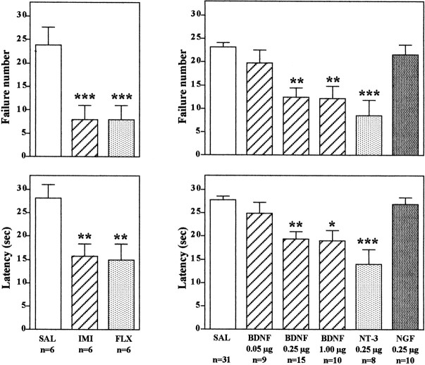
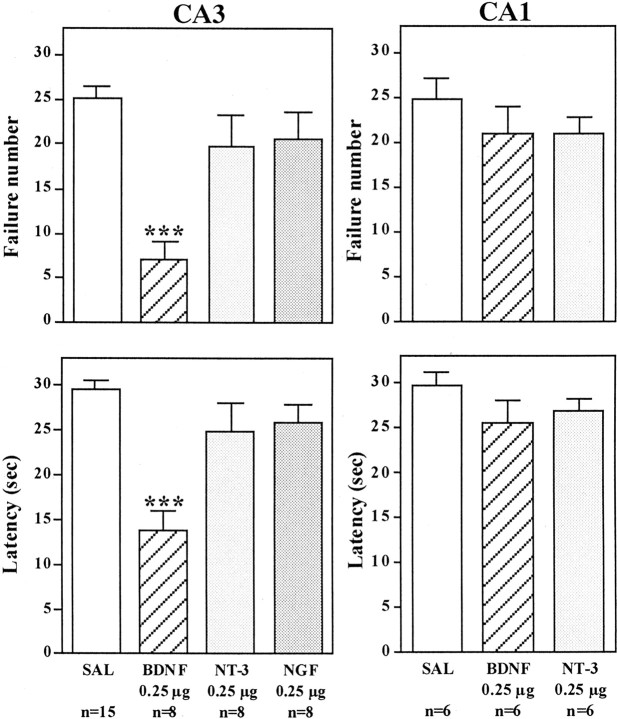
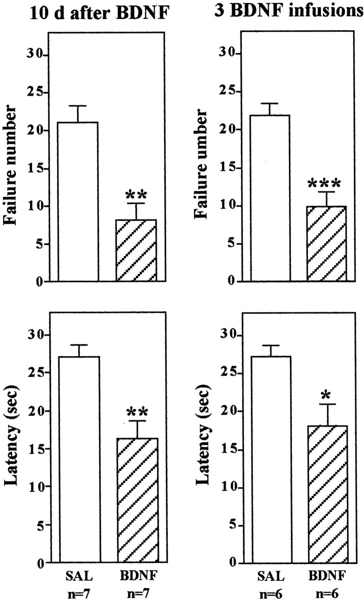
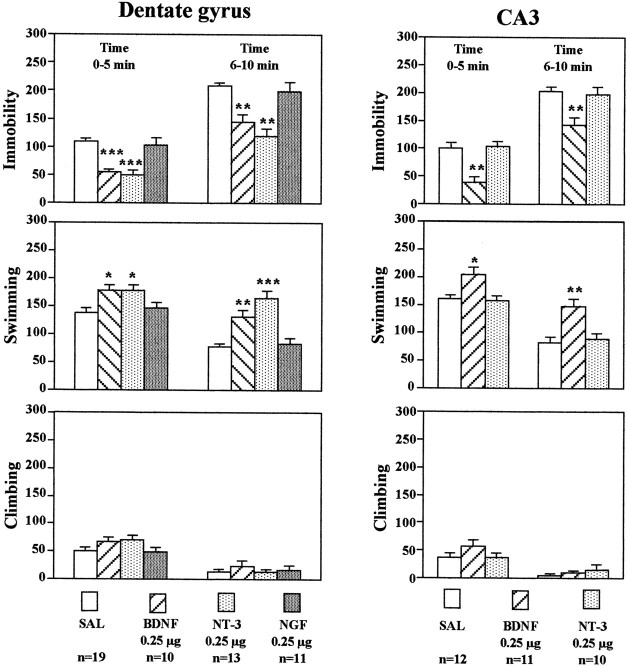
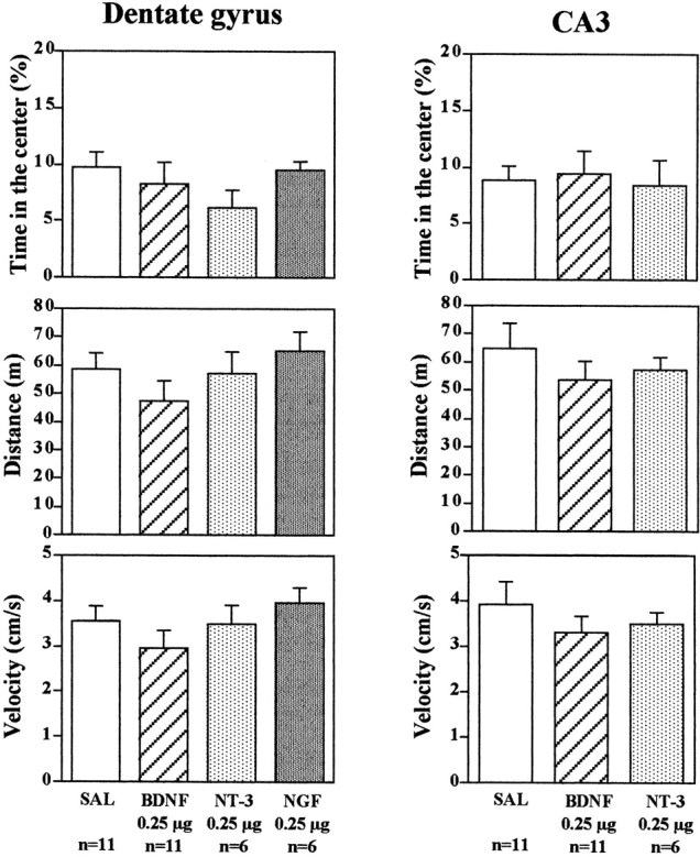
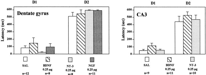
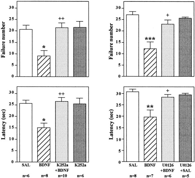
Previous studies demonstrated that antidepressant treatment increases the expression of brain-derived neurotrophic factor (BDNF) in rat hippocampus. The present study was conducted to test the hypothesis that BDNF in the hippocampus produces an antidepressant effect in behavioral models of depression, the learned helplessness (LH) and forced swim test (FST) paradigms. A single bilateral infusion of BDNF into the dentate gyrus of hippocampus produced an antidepressant effect in both the LH and FST that was comparable in magnitude with repeated systemic administration of a chemical antidepressant. These effects were observed as early as 3 d after a single infusion of BDNF and lasted for at least 10 d. Similar effects were observed with neurotrophin-3 (NT-3) but not nerve growth factor. Infusions of BDNF and NT-3 did not influence locomotor activity or passive avoidance. The results provide further support for the hypothesis that BDNF contributes to the therapeutic action of antidepressant treatment.


Figures

Fig. 1.
Fig. 1.
Infusion of BDNF into the dentate gyrus of hippocampus decreases escape failure in the LH paradigm. Animals were exposed to IES as described in Materials and Methods and then were administered the antidepressants or neurotrophic factors as indicated before conditioned avoidance testing. Imipramine (IMI) and fluoxetine (FLX) were administered for 7 d. BDNF, NT-3, NGF, or saline (SAL) was administered via bilateral infusion into the dentate gyrus at the doses indicated, and animals were tested in conditioned avoidance 3 d later. Escape failure and latency to escape were determined, and the results are expressed as mean ± SEM. The number of animals is listed under eachcolumn. *p < 0.05; **p < 0.01; ***p < 0.001 when compared with saline-injected controls (ANOVA and Scheffe's test).Left top, F(2,15) = 15.38; p = 0.0002; left bottom,F(2,15) = 9.282; p= 0.0024; right top,F(5,77) = 10.050; p< 0.0001; right bottom,F(5,77) = 10.872; p< 0.0001.
Fig. 2.
Fig. 2.
Influence of neurotrophic factor infusions into the CA3 and CA1 pyramidal cell layers on the LH paradigm. After exposure to IES, BDNF, NT-3, NGF, or saline (SAL) was infused into the CA3 or CA1 pyramidal cell layers of hippocampus, and conditioned avoidance was conducted 3 d later. Escape failure and latency to escape were determined, and the results are expressed as mean ± SEM. The number of animals is listed under each column. ***p < 0.001 when compared with saline-injected controls (ANOVA and Scheffe's test).Left top, F(3,33) = 12.667; p < 0.0001; left bottom,F(3,33) = 12.045; p< 0.0001; right top,F(2,15) = 0.824; p= 0.4575; right bottom,F(2,15) = 1.292; p= 0.3037.
Fig. 3.
Fig. 3.
BDNF regulation of LH behavior: long-term effects and multiple infusions. The LH paradigm was conducted as described in Materials and Methods, except conditioned avoidance testing was conducted 10 d after BDNF infusion into the dentate gyrus (left) or 3 d after infusions at three different rostrocaudal levels of dentate gyrus (right). The results are expressed as mean ± SEM. The number of animals is listed under each column. *p < 0.05; **p < 0.01; ***p < 0.001 when compared with saline-injected controls (Student's t test). Left top,F(1,12) = 4.172; p= 0.0013; left bottom,F(1,12) = 4.022; p= 0.0017; right top,F(1,10) = 4.824; p= 0.0007; right bottom,F(1,10) = 2.853; p= 0.0172.
Fig. 4.
Fig. 4.
Infusion of BDNF into the hippocampus has an antidepressant effect in the FST. BDNF, NT-3, NGF, or saline (SAL) was infused into the dentate gyrus or CA3 as indicated. Three days later, the durations of immobility, swimming, and climbing in the FST were determined. The results are divided into two 5 min time blocks as indicated and are the mean ± SEM of thenumber of animals indicated under eachcolumn. *p < 0.05; **p < 0.01; ***p < 0.001 when compared with the corresponding saline-injected controls (ANOVA and Scheffe's test). Left top, Immobility time, 0–5 min,F(3,49) = 16.348; p< 0.0001; immobility time, 6–10 min,F(3,49) = 15.823; p< 0.0001; left middle, swimming time, 0–5 min,F(3,49) = 4.997; p= 0.0042; swimming time, 6–10 min,F(3,49) = 18.025; p< 0.0001; left bottom, climbing time, 0–5 min,F(3,49) = 1.807; p= 0.1582; climbing time, 6–10 min,F(3,49) = 0.477; p= 0.7001; right top, immobility time, 0–5 min,F(2,30) = 15.749; p< 0.0001; immobility time, 6–10 min,F(2,30) = 8.219; p= 0.0014; right middle, swimming time, 0–5 min,F(2,30) = 6.811; p= 0.0036; swimming time, 6–10 min,F(2,30) = 10.129; p= 0.0004; right bottom, climbing time, 0–5 min,F(2,30) = 1.243; p= 0.3031; climbing time, 6–10 min,F(2,30) = 0.812; p= 0.4536. There were no significant treatment × time interactions.
Fig. 5.
Fig. 5.
Influence of neurotrophic factor infusion into the hippocampus on locomotor activity. BDNF, NT-3, NGF, or saline (SAL) was infused into the dentate gyrus or CA3, and 3 d later the times in center, distance, and velocity in an open field were determined. The results are the mean ± SEM of thenumber of animals indicated under eachcolumn. Left top, Time in the center,F(3,30) = 1.285; p= 0.2973; left middle, distance,F(3,30) = 1.143; p= 0.3479; left bottom, velocity,F(3,30) = 1.129; p= 0.3529; right top, time in the center,F(2,15) = 0.073; p= 0.9295; right middle, distance,F(2,15) = 0.650; p= 0.5359; right bottom, velocity,F(2,15) = 0.635; p= 0.5438.
Fig. 6.
Fig. 6.
Influence of neurotrophic factor infusions into hippocampus on passive avoidance. BDNF, NT-3, NGF, or saline (SAL) was infused into the dentate gyrus or CA3, and 3 d later, passive avoidance testing was conducted as described in Materials and Methods. The results are mean ± SEM of thenumber of animals indicated under eachcolumn. Left, Day 1 (D1),F(3,35) = 1.069; p= 0.3746; day 2 (D2),F(3,35) = 0.962; p= 0.4217; right, D1,F(2,27) = 1.980; p= 0.1577; D2, F(2,27) = 0.520; p = 0.6002.
Fig. 7.
Fig. 7.
BDNF immunohistochemistry after local infusion into the hippocampus. BDNF (0.25 or 1.0 μg) was infused into the dentate gyrus, and BDNF immunolabeling was determined at the time points indicated. Representative sections are shown for each time point and dose of BDNF. Arrows indicate the sites of infusion. Similar effects were observed in three or four separate animals for each condition.
Fig. 8.
Fig. 8.
Influence of BDNF infusion on levels of phospho-ERK immunolabeling. BDNF (0.25 μg) was infused into the dentate gyrus, and phospho-ERK immunolabeling was determined at the time points indicated. Representative low-power (left panels) and high-power (right panels) sections are shown for each time point. The locations of the dentate gyrus (DG) and CA3 and CA1 pyramidal cell layers (PyrCL) are indicated. Arrows indicate the infusion sites. Similar effects were observed in three or four separate animals for each condition.
Fig. 9.
Fig. 9.
Influence of BDNF infusion on levels of Fos immunolabeling. BDNF (0.25 μg) was infused into the dentate gyrus, and Fos immunolabeling was determined 4 hr later. Representative sections are shown, and similar effects were observed in three or four separate animals for each condition. The locations of the dentate gyrus (DG) and CA3 and CA1 pyramidal cell layers are indicated.
Fig. 10.
Fig. 10.
Infusion of K252a or U0126 blocks the effect of BDNF on conditioned avoidance. LH was conducted as in previous experiments; K252a was co-infused with BDNF, or U0126 was preinfused before BDNF into the dentate gyrus; and conditioned avoidance was conducted 3 d later. Escape failure and latency to escape were determined, and the results are expressed as mean ± SEM. Thenumber of animals is listed under eachcolumn. *p < 0.05; **p < 0.01; ***p < 0.001 when compared with saline-injected controls;+p < 0.05;++p < 0.01 when compared with the BDNF-injected group (ANOVA and Scheffe's test). Left top, F(3,26) = 7.586;p = 0.0008; left bottom,F(3,26) = 8.177; p= 0.0005; right top,F(3,22) = 10.804; p= 0.0001; right bottom,F(3,22) = 7.432; p= 0.0013.
Fig. 11.
Fig. 11.
Influence of U0126 on the induction of pERK in hippocampus. Saline or U0126 was infused 20 min before infusion of BDNF (0.25 μg) into the dentate gyrus. Phospho-ERK immunolabeling was determined at the time points indicated. Arrows indicate the sites of infusion. Representative sections are shown for each time point and condition. The locations of the dentate gyrus (DG) and CA3 and CA1 pyramidal cell layers (PyrCL) are indicated. Similar effects were observed in three or four separate animals for each condition.

References

    1. Altar C. Neurotrophins and depression. Trends Pharmacol Sci. 1999;20:59–61. - PubMed
    1. Amaral D, Witter MP. Hippocampal formulation. In: Paxinos G, editor. The rat nervous system: hippocampal formulation, Ed 2. Academic; San Diego: 1995.
    1. Bremner J, Narayan M, Anderson ER, Staib LH, Miller H, Charney DS. Smaller hippocampal volume in major depression. Am J Psychiatry. 2000;157:115–117. - PubMed
    1. Chang L, Karin M. Mammalian MAP kinase signalling cascades. Nature. 2001;410:37–40. - PubMed
    1. Chen A-H, Shirayama Y, Shin K-H, Neve RL, Duman RS. Expression of the cAMP response element binding protein (CREB) in hippocampus produces antidepressant effect. Biol Psychiatry. 2001;49:753–762. - PubMed

Publication types

MeSH terms