. 2002 Apr 16;99(8):5692-7.
doi: 10.1073/pnas.072097999. Epub 2002 Apr 9.

Somatosensory cortex dominated by the representation of teeth in the naked mole-rat brain

Affiliations

Somatosensory cortex dominated by the representation of teeth in the naked mole-rat brain

Kenneth C Catania et al. Proc Natl Acad Sci U S A. .

Abstract

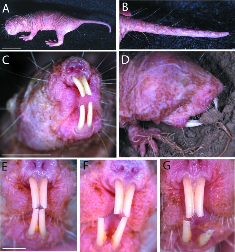
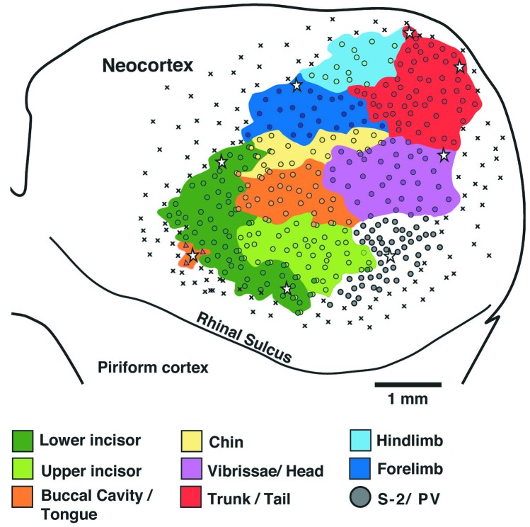
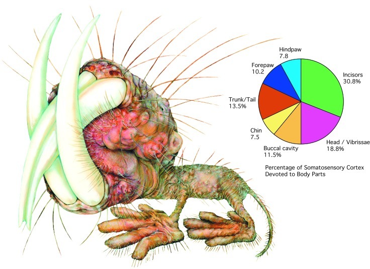
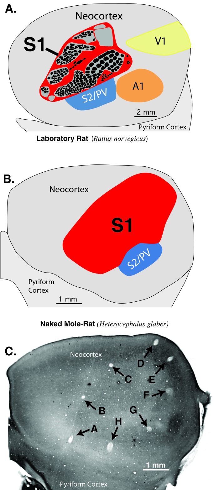
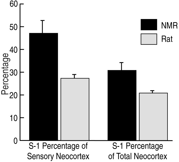
We investigated naked mole-rat somatosensory cortex to determine how brain areas are modified in mammals with unusual and extreme sensory specializations. Naked mole-rats (Heterocephalus glaber) have numerous anatomical specializations for a subterranean existence, including rows of sensory hairs along the body and tail, reduced eyes, and ears sensitive to low frequencies. However, chief among their adaptations are behaviorally important, enlarged incisors permanently exterior to the oral cavity that are used for digging, object manipulation, social interactions, and feeding. Here we report an extraordinary brain organization where nearly one-third (31%) of primary somatosensory cortex is devoted to the representations of the upper and lower incisors. In addition, somatosensory cortex is greatly enlarged (as a proportion of total neocortical area) compared with closely related laboratory rats. Finally, somatosensory cortex in naked mole-rats encompasses virtually all of the neocortex normally devoted to vision. These findings indicate that major cortical remodeling has occurred in naked mole-rats, paralleling the anatomical and behavioral specializations related to fossorial life.


Figures

Figure 1
Figure 1
The unusual anatomy of the naked mole-rat (H. glaber). (A) An adult mole-rat demonstrating the lack of fur and general proportions of the body. (B) The sensory hairs covering the tail that are used for navigation while traveling backward through tunnels. (C) Front view of a naked mole-rat showing the enlarged incisors that are located permanently exterior to the oral cavity. Note also the sensory whiskers covering the front part of the face and mouth region. (D) A mole-rat moves a small piece of earth with the teeth. (EG) A remarkable feature of the lower incisors is their ability to move independently. This is possible because of a flexible mandibular symphysis connecting the two halves of the lower jaw. The specimen shown here was anesthetized to document their mobility in the dorso-ventral and medio-lateral planes. Analysis of slow-motion videotaped behavior shows that independent movements in these different directions are common. (Scales bars: 2 cm in A; 1 cm in C; 5 mm in E.)
Figure 2
Figure 2
Results of microelectrode recordings from naked mole-rat case 4 showing the extent of S1 and the location of the representations for different body parts in the left hemisphere. Each circle marks an electrode penetration where neurons responded to tactile stimulation of the body surface. Each colored region indicates the extent of the representation of a different body part. The representation of the dentition (Green) was found to be greatly expanded in naked mole-rats, taking up approximately 31% of S1. Note also the relatively large size of S1 overall, and its extension into far caudal and medial cortex where visual cortex is usually located in other mammals. ⋆, microlesions; ×, unresponsive sites. Medial is up and rostral is to the left.
Figure 3
Figure 3
The relative sizes of different sensory representations in naked mole-rats S1. The chart on the right shows the percentage of cortex devoted to different body parts. On the left the different body parts are illustrated according to their cortical proportions. This “mole-ratunculus” provides a graphic illustration of the cortical magnification of the incisors and head (illustration by Lana Finch).
Figure 4
Figure 4
A comparison of the size and location of primary somatosensory cortex between laboratory rats (Long–Evans) and the naked mole-rat. (A) The location and size of S1 in a rat drawn from a section of layer 4 cortex processed for cytochrome oxidase histochemistry. Black ovals within S1 reflect the locations of cytochrome oxidase dark modules that represent whiskers and skin surfaces (30). S1 takes up approximately 21% of the neocortex in rats and is located rostral to primary visual (V1) and auditory cortex (A1). (B) The location and size of S1 in the naked mole-rat. S1 takes up approximately 31% of neocortex and extends far caudal and medial, occupying cortical territory usually taken up by visual areas in other mammals. (C) A section of flattened cortex from a naked mole-rat showing microlesions made where neurons responded to mechanosensory stimulation of body parts and outlining much of S1. Note the caudo-medial location of lesions D and E. Neurons at the lesions responded in the following sequence: A, tongue; B, lower incisor; C, forelimb; D and E, tail; F, facial vibrissae; G, hindlimb (in S2/PV); H, lower incisor.
Figure 5
Figure 5
Comparison of the overall proportion of neocortex taken up by S1 in the naked mole-rat and Long–Evans laboratory rat. S1 was approximately 50% larger, as a proportion of total neocortex, in the naked mole-rat (Right). A second comparison is the proportion of sensory cortex taken up by S1 in the two species (Left). Total sensory cortex here was defined as cortex caudal to the rostral border of S1, which necessarily includes all visual cortex (V1 and V2), auditory cortex, and the secondary somatosensory area (S2) and parietal ventral somatosensory area (PV). Because mole-rats have less sensory cortex overall, S1 takes up a larger proportion of neocortex devoted to all sensory areas. Bars indicate SD.

References

    1. Jarvis J U M. Science. 1981;212:571–573. - PubMed
    1. Jarvis J U M, O'Riain M J N, Bennett C, Shermman P W. Trends Ecol Evol. 1994;9:47–51. - PubMed
    1. O'Riain M J, Jarvis J U, Faulkes C G. Nature (London) 1996;380:619–621. - PubMed
    1. O'Riain M J, Jarvis J U, Alexander R, Buffenstein R, Peeters C. Proc Natl Acad Sci USA. 2000;97:13194–13197. - PMC - PubMed
    1. Faulkes C G, Bennett N C. Trends Ecol Evol. 2001;16:184–190. - PubMed

Publication types

LinkOut - more resources