. 2002 Apr 16;99(8):5655-60.
doi: 10.1073/pnas.072557799. Epub 2002 Apr 9.

Activation of mislocalized opsin kills rod cells: a novel mechanism for rod cell death in retinal disease

Affiliations

Activation of mislocalized opsin kills rod cells: a novel mechanism for rod cell death in retinal disease

Peter D Alfinito et al. Proc Natl Acad Sci U S A. .

Abstract

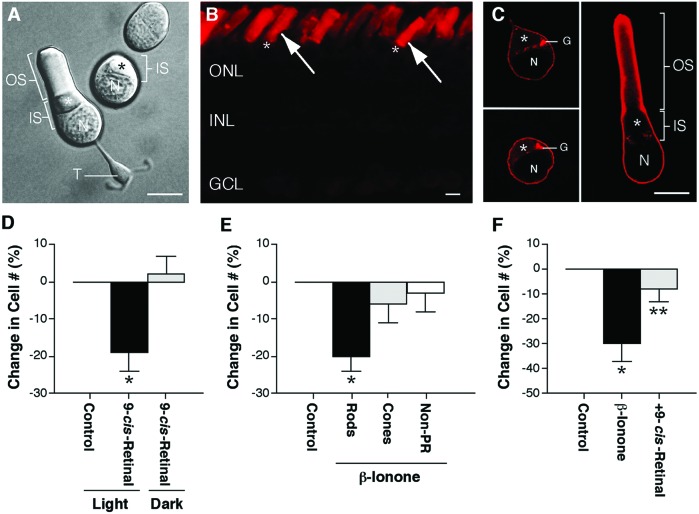
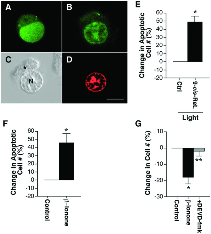
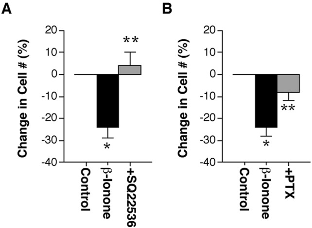
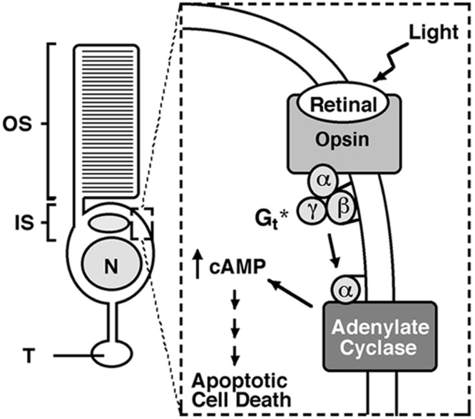
Rod photoreceptors are highly compartmentalized sensory neurons that maintain strict ultrastructural and molecular polarity. Structural subdivisions include the outer segment, inner segment, cell body, and synaptic terminal. The visual pigment rhodopsin is found predominantly in membranes of the rod cell outer segment but becomes mislocalized, appearing throughout the plasma membrane of the cell in many retinal diseases and injuries. Currently, there is no known link between rhodopsin redistribution and rod cell death. We propose that activation of mislocalized rhodopsin kills rod cells by stimulating normally inaccessible signaling pathways. This hypothesis was tested in primary retinal cell cultures, which contain photoreceptors. In rod photoreceptors, opsin immunofluorescence occurred throughout the rod cell plasma membrane. Activation of this mislocalized opsin by photostimulation after formation of isorhodopsin or by incubation with beta-ionone (opsin agonist) killed 19-30% of rod cells. Rod cell death was apoptotic, as indicated by marked chromatin condensation and the requirement for caspase-3 activation. Rod cell death could be induced by forskolin (adenylate cyclase agonist), and conversely, beta-ionone-induced cell death could be blocked by cotreatment with SQ22536 (an adenylate cyclase inhibitor). Pertussis toxin (a G protein inhibitor) also blocked beta-ionone-induced cell death. The data support a mechanism by which activation of mislocalized opsin initiates apoptotic rod cell death through G protein stimulation of adenylate cyclase.


Figures

Figure 1
Figure 1
Activation of mislocalized opsin kills rod cells. (A) Structural subdivisions of rod cells viewed with Nomarski optics. Rod cells in culture exist with or without their outer segment. The ellipsoid (*), an accumulation of mitochondria, lies within the inner segment. OS, outer segment; IS, inner segment; N, nucleus; T, synaptic terminal. (Bar = 10 μm.) [Reproduced from ref. , p. 111 by courtesy of Marcel Dekker, Inc.)]. (B) Conventional fluorescence image of a 10-μm frozen section of the salamander retina labeled with a monoclonal antibody to rod opsin (4D2) and a rhodamine-conjugated secondary antibody. Intense opsin staining was present in rod cell outer segments (arrows) but not in inner segments indicated by (*) or cell bodies, located in the ONL. ONL, outer nuclear layer; INL, inner nuclear layer; GCL, ganglion cell layer. (Bar = 10 μm.) (C) One-μm optical sections of isolated rod cells obtained by confocal microscopy showed opsin labeling throughout the plasma membrane of the inner segment and cell body. Opsin labeling also was seen within the Golgi apparatus. OS, outer segment; IS, inner segment; N, nucleus; G, Golgi apparatus; *, ellipsoid. (Bar = 10 μm.) (D) Percent change in rod cell number after photoactivation of reconstituted isorhodopsin. Light plus 10 μM 9-cis-retinal killed 19% of rod cells in culture compared with light-treated phospholipid controls. When maintained in the dark, rod cell number was unchanged by 9-cis-retinal treatment. In n = 9 experiments from three animals, a total of 1,874 rod cells was counted; *, P < 0.01, means ± SEM. (E) Percent change in cell number after treatment with 2.5 μM β-ionone. Rod cell number decreased 20% in β-ionone-treated cultures compared with ethanol controls. Cone and nonphotoreceptor cell numbers did not change. In n = 6 experiments from two animals, a total of 1,425 rod, 1,335 cone, and 3,735 nonphotoreceptor cells was counted; *, P < 0.01, means ± SEM. (F) The effect of β-ionone on rod cell death was blocked by cotreatment with 10 μM 9-cis-retinal in the dark. In n = 5 experiments from two animals, a total of 671 rod cells was counted; *, P < 0.02 compared with control; **, P < 0.05 compared with β-ionone treated; unpaired t test, means ± SEM.
Figure 2
Figure 2
Characterization of rod cell death. (A, B, D) One-μm optical sections of isolated rod cells fluorescently labeled with AO and EtdBr obtained by confocal microscopy. (C) Nomarski optics image of an isolated rod cell treated with high K+ Ringer's solution demonstrating rod cell morphology after loss of membrane integrity. (A) Live rod cells had green (AO), homogeneously stained chromatin. (B) Early apoptotic rod cells had green (AO), condensed chromatin. (C) Rod cells that had lost membrane integrity could be identified by their rectangular, elongated ellipsoid. *, ellipsoid; N, nucleus. (D) Late apoptotic cells with permeant membranes had red EtdBr, condensed chromatin. (Bar = 10 μm.) (E) Percent change in the number of apoptotic rod cells after treatment with light and 10 μM 9-cis-retinal. Photoactivation of reconstituted isorhodopsin increased apoptotic rod cell death by 49%. In n = 5 experiments from two animals, a total of 83 apoptotic rod cells was counted; *, P < 0.01, means ± SEM. (F) Percent change in the number of apoptotic rod cells after treatment with 2.5 μM β-ionone. β-ionone treatment increased apoptotic rod cell death by 46%. In n = 9 experiments from three animals, a total of 120 apoptotic rod cells was counted; *, P < 0.01, means ± SEM. (G) The effect of β-ionone on rod cell death was blocked by preincubation and cotreatment with 1 μM DEVD-fmk. In n = 11 experiments from four animals, a total of 4,417 rod cells was counted; *, P < 0.01 compared with control; **, P < 0.01 compared with β-ionone treated; unpaired t test, means ± SEM.
Figure 3
Figure 3
Mislocalized opsin activity stimulates normally inaccessible signaling pathways. (A) Inhibition of adenylate cyclase with 100 μM SQ22536 blocked β-ionone-induced cell death. In n = 6 experiments from two animals, a total of 855 rod cells was counted in 20 fields at 20×; *, P < 0.01 compared to control; **, P < 0.005 compared with β-ionone treated; unpaired t test, means ± SEM. (B) Preincubation and cotreatment with 100 ng/ml of pertussis toxin blocked β-ionone-induced rod cell death. In n = 6 experiments from two animals, a total of 1,226 rod cells was counted in 40 fields at 40×; *, P < 0.01 compared with control; **, P < 0.03 compared with β-ionone treated; unpaired t test, means ± SEM.
Figure 4
Figure 4
Proposed mechanism of rod cell death. During retinal degenerative disease, opsin becomes mislocalized to the plasma membrane of the inner segment, cell body, and synaptic terminal. Photostimulation of mislocalized rhodopsin activates G proteins such as transducin. In the absence of its normal target, PDE, transducin binds to and activates adenylate cyclase. The subsequent increase in cAMP levels initiates signals within the rod cell that eventually lead to caspase-3 activation and apoptotic cell death. *, photoactivation of mislocalized opsin may kill rod cells through the stimulation of other G proteins. OS, outer segment; IS, inner segment; N, nucleus; T, synaptic terminal.

References

    1. Schmidt S Y. In: Handbook of Neurochemistry, Volume 10. Lajthe A, editor. New York: Plenum; 1985. pp. 461–507.
    1. Li Z-Y, Milam A H. In: Degenerative Diseases of the Retina. Anderson R E, editor. New York: Plenum; 1995. pp. 1–8.
    1. Portera-Cailliau C, Sung C-H, Nathans J, Adler R. Proc Natl Acad Sci USA. 1994;91:974–978. - PMC - PubMed
    1. Cook B, Lewis G P, Fisher S K, Adler R. Invest Ophthalmol Visual Sci. 1995;36:990–996. - PubMed
    1. Abler A S, Chang C J, Ful J, Tso M O M, Lam T T. Res Commun Mol Pathol Pharmacol. 1996;92:177–189. - PubMed

Publication types

MeSH terms