Comparative Study
. 2002 May;184(9):2473-80.
doi: 10.1128/JB.184.9.2473-2480.2002.

Firm but slippery attachment of Deinococcus geothermalis

Affiliations
Comparative Study

Firm but slippery attachment of Deinococcus geothermalis

M Kolari et al. J Bacteriol. 2002 May.

Abstract

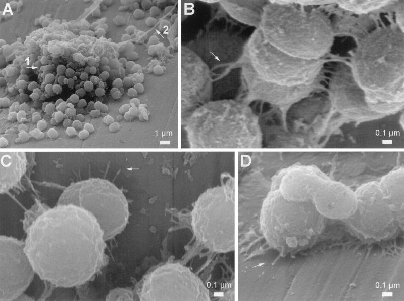
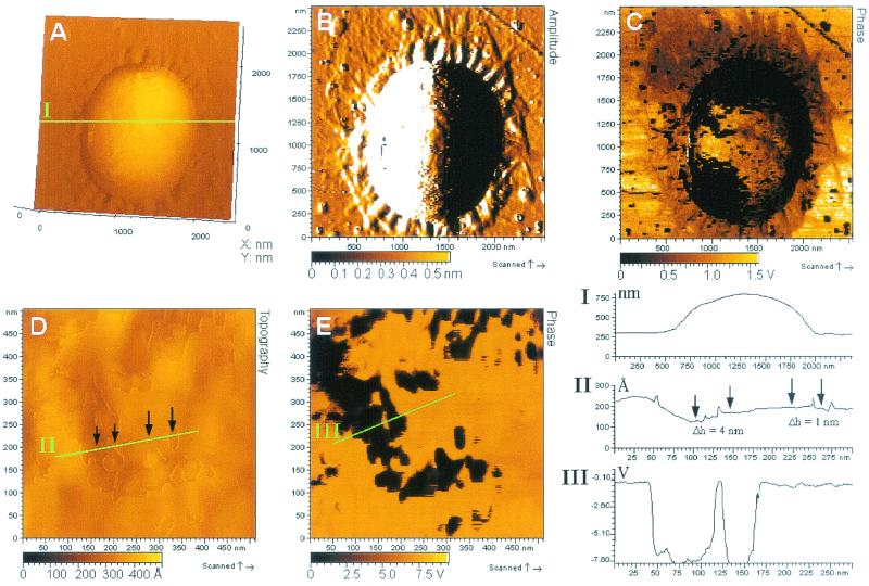
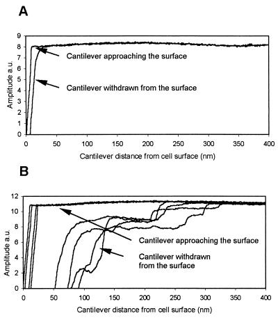
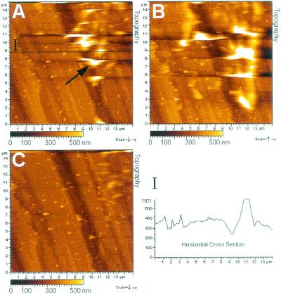
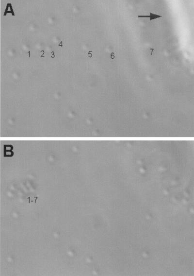
Bacterial biofilms impair the operation of many industrial processes. Deinococcus geothermalis is efficient primary biofilm former in paper machine water, functioning as an adhesion platform for secondary biofilm bacteria. It produces thick biofilms on various abiotic surfaces, but the mechanism of attachment is not known. High-resolution field-emission scanning electron microscopy and atomic force microscopy (AFM) showed peritrichous adhesion threads mediating the attachment of D. geothermalis E50051 to stainless steel and glass surfaces and cell-to-cell attachment, irrespective of the growth medium. Extensive slime matrix was absent from the D. geothermalis E50051 biofilms. AFM of the attached cells revealed regions on the cell surface with different topography, viscoelasticity, and adhesiveness, possibly representing different surface layers that were patchily exposed. We used oscillating probe techniques to keep the tip-biofilm interactions as small as possible. In spite of this, AFM imaging of living D. geothermalis E50051 biofilms in water resulted in repositioning but not in detachment of the surface-attached cells. The irreversibly attached cells did not detach when pushed with a glass capillary but escaped the mechanical force by sliding along the surface. Air drying eliminated the flexibility of attachment, but it resumed after reimmersion in water. Biofilms were evaluated for their strength of attachment. D. geothermalis E50051 persisted 1 h of washing with 0.2% NaOH or 0.5% sodium dodecyl sulfate, in contrast to biofilms of Burkholderia cepacia F28L1 or the well-characterized biofilm former Staphylococcus epidermidis O-47. Deinococcus radiodurans strain DSM 20539(T) also formed tenacious biofilms. This paper shows that D. geothermalis has firm but laterally slippery attachment not reported before for a nonmotile species.


Figures

FIG. 1.
FIG. 1.
FESEM analysis of D. geothermalis E50051 biofilms grown on polished (1,000 grit) stainless steel. Laboratory biofilms were grown in sterilized paper machine circulating water medium (1 day, 45°C, 160 rpm). The few rod-shaped dead bacteria and cellulose fibrils adhering to the growing deinococcal colonies (arrows 1 and 2 in panel A) originated from the heat-sterilized medium. Thin adhesion threads mediated the cell-to-cell attachment and connected the cells to the stainless steel surface (arrows in panels B, C, and D).
FIG. 2.
FIG. 2.
AFM analysis of D. geothermalis E50051 cells attached on a glass surface. The biofilm was grown in R2 broth (1 day, 45°C, 160 rpm), rinsed with water, allowed to air dry for 15 min, and imaged in the AAC mode. (A, B, and C) Topography, amplitude, and phase images, respectively, of one attached cell. (D and E) Close-up topography and phase images, respectively, of the surface of the same cell. Green lines indicate the cut positions of the horizontal cross sections I, II, and III.
FIG. 3.
FIG. 3.
Amplitude-versus-distance curves representing the forces acting on the AFM tip as it approached and was withdrawn from the bright (A) and dark (B) areas of the cell surface of D. geothermalis E50051 visible in the AFM phase image (Fig. 2C). The approach of the tip towards the cell surface resulted in a similar amplitude change in both areas, whereas during withdrawal from the dark surface areas (panel B, four example curves) the tip experienced attractive forces, which retarded the return to the free oscillation amplitude. a.u., arbitrary units.
FIG. 4.
FIG. 4.
AFM analysis of D. geothermalis E50051 cells attached to stainless steel. The biofilm was grown in R2 broth (1 day, 45°C, 160 rpm) and imaged in the MAC mode in water. The cells are seen as white spots like the one indicated by an arrow in the topography image in panel A. Cross section I confirms the white spots as deinococcal cells. The images in panels A, B, and C are from three subsequent scannings at the same location and show that one scan repositioned the live cells by 1 to 5 μm (panel A versus panel B) but did not detach these cells from the surface. The next scan caused sliding of the cells outside the scan area (C).
FIG. 5.
FIG. 5.
Phase-contrast light microscopy images of D. geothermalis E50051 cells attached on glass surface. (A) Positions of seven numbered cells on the surface (the arrow points to shadow of a glass capillary). (B) New positions of the same cells 1 to 7 after being pushed with the tip of the glass capillary. All seven pushed cells repositioned with no detachment from the surface, indicating a mechanism of slippery attachment. Panel B also shows that all untouched cells retained their positions. More images are shown at http://www.honeybee.helsinki.fi/users/mkolari/deino.html.

References

    1. Auerbach, I. D., C. Sorensen, H. G. Hansma, and P. A. Holden. 2000. Physical morphology and surface properties of unsaturated Pseudomonas putida biofilms. J. Bacteriol. 182:3809-3815. - PMC - PubMed
    1. Beech, I. B. 1996. The potential use of atomic force microscopy for studying corrosion of metals in the presence of bacterial biofilms. Int. Biodeterior. Biodegrad. 37:141-149.
    1. Beech, I. B., C. W. S. Cheung, D. B. Johnson, and J. R. Smith. 1996. Comparative studies of bacterial biofilms on steel surfaces using atomic force microscopy and environmental scanning electron microscopy. Biofouling 10:65-77. - PubMed
    1. Blanco, M. A., C. Negro, I. Gaspar, and J. Tijero. 1996. Slime problems in the paper and board industry. Appl. Microbiol. Biotechnol. 46:203-208.
    1. Bremer, P. J., G. G. Geesey, and B. Drake. 1992. Atomic force microscopy examination of the topography of a hydrated bacterial biofilm on a copper surface. Curr. Microbiol. 24:223-230.

Publication types

MeSH terms