

Case Reports
Angiographic and embryologic considerations in five cases of middle cerebral artery fenestration
Affiliations
- PMID: 11950648
- PMCID: PMC7975097
Item in Clipboard
Case Reports
Angiographic and embryologic considerations in five cases of middle cerebral artery fenestration
AJNR Am J Neuroradiol.
2002 Apr.
Abstract
Five cases of unilateral middle cerebral artery fenestration were observed during the prospective evaluation of 1466 consecutive cerebral angiograms (0.43% of 1170 patients) between January 1999 and July 2001. In each case, an early branching temporopolar artery was seen to arise from the inferior limb of the fenestrated segment. This finding suggests that early branching temporopolar arteries may participate in the formation of middle cerebral artery fenestration by interfering with the normal fetal development of the middle cerebral artery.
Figures
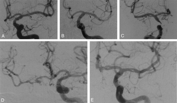
Five angiographic observations of MCA fenestration. Anteroposterior digital subtraction angiograms. In each case, the fenestration is located in the M1 segment of the MCA. A, Case 1. Fenestration of the left MCA in a 75-year-old woman. The arrowhead indicates an early branching TPA; open arrow, prominent lenticulostriate artery arising from superior aspect of fenestrated segment. B, Case 2. Fenestration of the left MCA in a 72-year-old man. The arrowhead indicates an early branching TPA; open arrow, prominent lenticulostriate artery arising from superior aspect of fenestrated segment. C, Case 3. Fenestration of the right MCA in a 39-year-old man. The arrowhead indicates an early branching TPA. D, Case 4. Fenestration of the right MCA in an 82-year-old woman. E, Case 5. Fenestration of the left MCA in a 50-year-old woman.
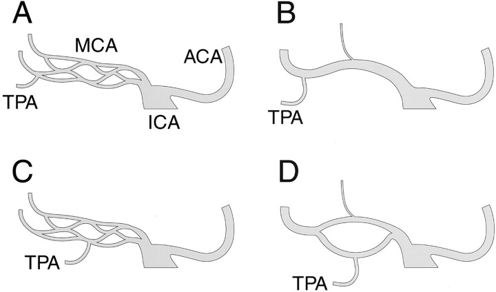
Hypothetic mechanism underlying MCA fenestration formation. Anteroposterior schematic representations of the ICA bifurcation into anterior cerebral arteries (ACA) and MCA. A, Normal MCA development, fetal stage. At this stage, the MCA is constituted by multiple arterial twigs arising from the distal ICA. B, Normal MCA development, adult configuration. The multiple MCA twigs have been replaced by a single proximal MCA trunk (M1 segment). C, Fenestrated MCA development, fetal stage. The fetal stage is similar to the normal development illustrated in A. An early branching TPA is shown, which might predispose to the formation of fenestration, either by precluding the fusion of several primitive twigs into a single trunk or by interfering with the regression of some of the twigs. D, Fenestrated MCA development, adult configuration. Fenestration of the M1 segment is shown, with an early branching TPA arising from the inferior aspect of the fenestrated segment, as observed in our five angiographic cases.
References
-
- Umansky F, Dujovny M, Ausman JI, Diaz FG, Mirchandani HG. Anomalies and variations of the middle cerebral artery: a microanatomical study. Neurosurgery 1988;22:1023–1027 - PubMed
-
- Hassler O. Morphological studies of the large cerebral arteries. Acta Psychiatr Neurol Scand 1961;36([suppl 154]):37–42 - PubMed
-
- Gibo H, Lenkey C, Rhoton AL. Microsurgical anatomy of the supraclinoid portion of the internal carotid artery. Neurosurgery 1981;55:560–574 - PubMed
-
- Ito J, Maeda H, Inoue K, Onishi Y. Fenestration of the middle cerebral artery. Neuroradiology 1977;13:37–39 - PubMed
Publication types
MeSH terms
LinkOut - more resources
Full Text Sources