. 2002 Apr;23(4):635-43.

Imaging findings of cochlear nerve deficiency

Affiliations

Imaging findings of cochlear nerve deficiency

Christine M Glastonbury et al. AJNR Am J Neuroradiol. 2002 Apr.

Abstract

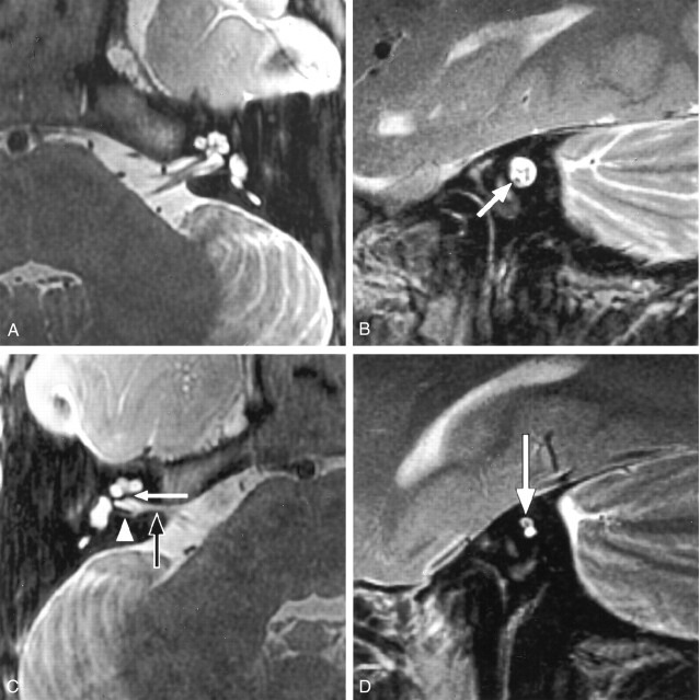
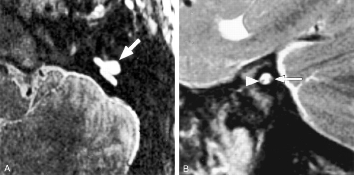
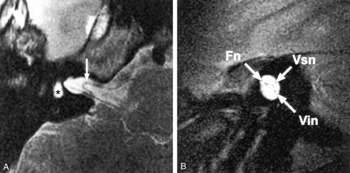
Background and purpose: High-resolution T2-weighted fast spin-echo MR imaging provides excellent depiction of the cisternal and intracanalicular segments of the vestibulocochlear and facial nerves. Absence or reduction in caliber of the cochlear nerve (deficiency) has been described in association with congenital sensorineural hearing loss (SNHL). Depiction of cochlear nerve integrity may be important for diagnosis and management of SNHL.

Methods: We retrospectively reviewed high-resolution T2-weighted fast spin-echo MR images of 22 patients examined for SNHL who had deficiency of the cochlear nerve. Images were evaluated for the presence and comparative size of the component nerves (facial, cochlear, superior vestibular, and inferior vestibular nerves), relative size of the internal auditory canal (IAC), and any associated inner ear abnormalities. The clinical history, results of the clinical examination, and audiometric findings were reviewed for each patient.

Results: Deficiency of the cochlear nerve was observed in 12 patients with congenital SNHL and in 10 patients with acquired SNHL. Hypoplasia of the IAC was observed in association with congenital deficiency of the cochlear nerve in 11 of 12 patients. Deficiency of the cochlear nerve was observed in association with acoustic schwannoma in two cases and with acquired labyrinthine abnormalities in seven cases. Hypoplasia of the IAC was not observed in association with acquired SNHL.

Conclusion: Deficiency of the cochlear nerve can be shown by high-resolution T2-weighted fast spin-echo MR imaging. Deficiency may be observed in association with congenital or acquired SNHL and may be important in the assessment of patients for cochlear implantation. Hypoplasia of the IAC is an indicator of congenital cochlear nerve deficiency.


Figures

F<sc>ig</sc> 1.
Fig 1.
High-resolution T2-weighted fast spin-echo MR images provide excellent depiction of the cisternal and intracanalicular segments of the facial nerve and the three divisions of the vestibulocochlear nerve. A, Axial view of the left cerebellopontine angle and IAC shows the normal anatomy. Parallel lines illustrate the plane prescribed for oblique plane sagittal images obtained perpendicular to the nerves of the IAC. B, Oblique plane sagittal image obtained at the fundus of the IAC, oriented with anterior to the left and the cerebellum to the right. High-signal-intensity CSF delineates the four nerves of the IAC. In the anterior aspect of the canal, the facial nerve (Fn) lies superiorly, with the cochlear nerve (Cn) inferior to it. Superior (Vsn) and inferior (Vin) vestibular nerves lie posteriorly.
F<sc>ig</sc> 2.
Fig 2.
Congenital absence of the cochlear nerve with an isolated cochlea. Axial and oblique sagittal T2-weighted fast spin-echo MR images of a 5-year-old girl with profound unilateral hearing loss (patient C8). A, Image of the normal left side shows the normal contours of the cochlea and other labyrinthine structures. B, IAC is of normal size and contains four nerves of comparative size. Cochlear nerve lies anteroinferiorly (arrow). C, Right side shows a deformed contour of the IAC (black arrow). Low-signal-intensity bar separates the fundus of the IAC from the modiolus (white arrow), which was confirmed to be bony at CT. We describe this as an isolated cochlea. The arrowhead indicates a singular canal containing the nerve of the posterior semicircular canal. D, Oblique sagittal image of the distal IAC shows a solitary nerve within the superior aspect of the small, deformed canal (arrow). The cochlear nerve is absent in this patient with normal facial nerve function.
F<sc>ig</sc> 3.
Fig 3.
Bilateral cochlear nerve absence in a 7-year-old patient with severe dysplasia and bilateral absence of the cochlea (patient C10). Facial nerve function was preserved. A, Axial T2-weighted fast spin-echo MR image of the left side shows dysplasia of the vestibule and semicircular canals (arrow). Cochlea was absent. B, Oblique sagittal MR image obtained on the same side shows a small IAC containing only two nerves. The intact facial nerve lies anteriorly (arrowhead). We suspected that the posterior nerve was vestibular, on the basis of its position and the absence of cochlear structures (arrow).
F<sc>ig</sc> 4.
Fig 4.
Deficient cochlear nerve in a 60-year-old patient with a 50-year history of left hearing loss after a motor vehicle accident (patient A10). This patient presented with a 3-month history of right-sided hearing loss. A, Axial T2-weighted fast spin-echo MR image shows a small left modiolus (arrow) but no other structural cochlear abnormalities. B, Corresponding oblique sagittal image shows a small caliber cochlear nerve (arrow) in a normal-sized IAC. C, Axial T2-weighted fast spin-echo MR image of the right side shows no abnormality to explain the recent hearing loss. D, Oblique sagittal T2-weighted fast spin-echo MR image of the right side also shows no abnormality to explain the recent hearing loss. A normal-caliber cochlear nerve is seen (arrow). Cochlear implantation was successfully performed on this side.
F<sc>ig</sc> 5.
Fig 5.
Acquired cochlear nerve deficiency in a 14-year-old patient with hearing loss after meningococcal meningitis at the age of 6 months (patient A3). Bilateral labyrinthitis ossificans was shown by CT (not shown). A, Axial T2-weighted fast spin-echo MR image of the right side shows loss of the normal high-signal-intensity CSF in the labyrinth. The asterisk indicates the vestibule; arrow, arterior inferior cerebellar artery (AICA) loop. B, Corresponding oblique sagittal image shows three nerves in the IAC. The cochlear nerve cannot be identified. Note the normal size of the IAC in this acquired cochlear nerve deficiency. Fn indicates the facial nerve; Vsn, superior vestibular nerve; and Vin, inferior vestibular nerve.
F<sc>ig</sc> 6.
Fig 6.
Acquired left-sided hearing loss of unknown etiology in patient A6. Axial high-resolution MR images showed a small left modiolus but no other labyrinthine abnormality (not shown). Cn indicates cochlear nerve; Fn indicates facial nerve; Vin, inferior vestibular nerve; and Vsn, superior vestibular nerve. A, Right oblique sagittal T2-weighted fast spin-echo MR image clearly shows four normally sized nerves in the IAC. B, On the affected left side, the cochlear nerve is not identified. Note the normal size of the IAC, which indicates an acquired abnormality after in utero formation of the canal.

References

    1. Rubinstein D, Sandberg EJ, Cajade-Law AG. Anatomy of the facial and vestibulocochlear nerves in the internal auditory canal. AJNR Am J Neuroradiol 1996;17:1099–1105 - PMC - PubMed
    1. Kim HS, Kim DI, Chung IH, Lee WS, Kim KY. Topographical relationship of the facial and vestibulocochlear nerves in the subarachnoid space and internal auditory canal. AJNR Am J Neuroradiol 1998;19:1155–1161 - PMC - PubMed
    1. Casselman JW, Offeciers FE, Govaerts PJ, et al. Aplasia and hypoplasia of the vestibulocochlear nerve: diagnosis with MR imaging. Radiology 1997;202:773–781 - PubMed
    1. Nadol JB Jr, Xu WZ. Diameter of the cochlear nerve in deaf humans: implications for cochlear implantation. Ann Otol Rhinol Laryngol 1992;101:988–993 - PubMed
    1. Felix H, Hoffmann V. Light and electron microscopic investigation of cochlear nerve specimens from profoundly deaf patients. Acta Otolaryngol Suppl 1985;423:67–72 - PubMed