. 2002 May;50(5):658-64.
doi: 10.1136/gut.50.5.658.

Characteristics of the peroxisome proliferator activated receptor gamma (PPARgamma) ligand induced apoptosis in colon cancer cells

Affiliations

Characteristics of the peroxisome proliferator activated receptor gamma (PPARgamma) ligand induced apoptosis in colon cancer cells

T Shimada et al. Gut. 2002 May.

Abstract

Background: Involvement of peroxisome proliferator activated receptor gamma (PPARgamma) in the growth response of colon cancer cells has been suggested.

Aims: To investigate the characteristics of PPARgamma induced apoptosis in colon cancer cells.

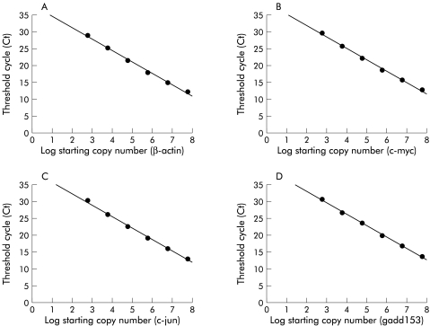
Methods: The effects of ligands for each of the PPAR subtypes (alpha, delta, and gamma) on DNA synthesis and cell viability were examined in HT-29 colon cancer cells. Modulation of apoptosis related gene expression by PPARgamma ligands was screened with cDNA arrays, and the results were confirmed by quantitative reverse transcription-polymerase chain reaction (RT-PCR) analysis.

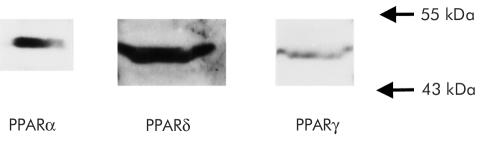
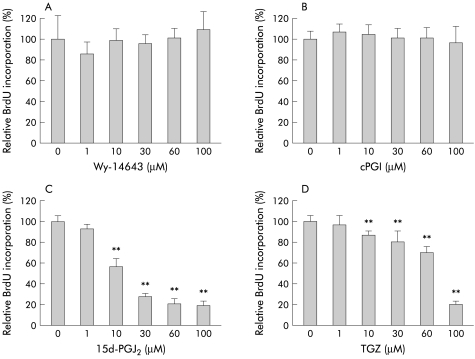
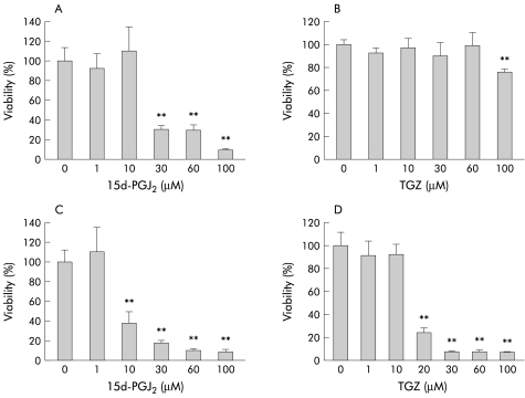
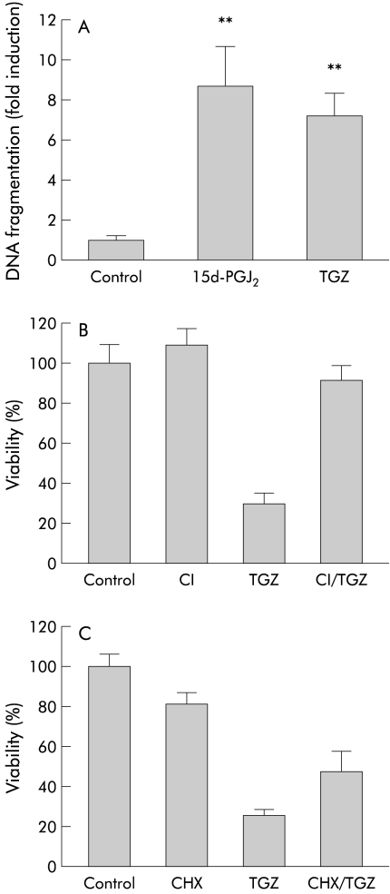
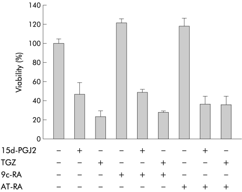
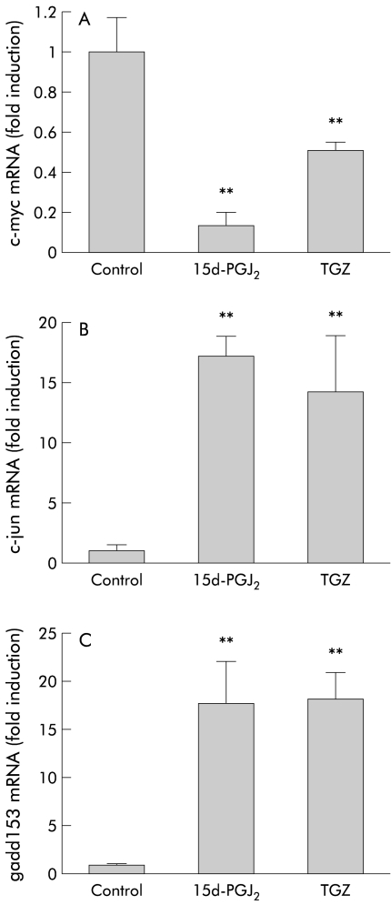
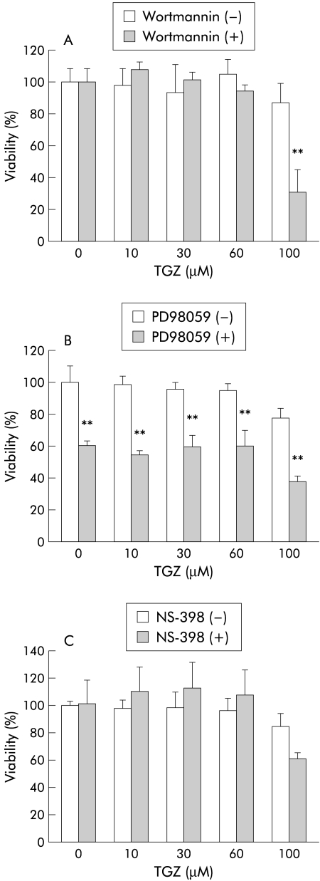
Results: PPARalpha, PPARdelta, and PPARgamma were all expressed in HT-29 cells. PPARgamma ligands, 15-deoxy-delta(12,)(14)-prostaglandin J2 (15d-PGJ2) and troglitazone (TGZ), suppressed DNA synthesis of HT-29 cells whereas ligands for PPARalpha and PPARdelta had no significant effects. Both 15d-PGJ2 and TGZ induced HT-29 cell death in a dose dependent manner which was associated with an increase in fragmented DNA and was sensitive to a caspase inhibitor. Among several genes selected by cDNA array screening, quantitative RT-PCR analysis confirmed downregulation of c-myc expression and upregulation of c-jun and gadd153 expression by 15d-PGJ2 and TGZ. PPARgamma induced apoptosis was antagonised by the presence of serum in the culture medium, and interaction between PPARgamma signalling and cell survival signalling through the phosphatidylinositol 3-kinase pathway was suggested.

Conclusions: As c-myc is an important target gene of the adenomatous polyposis coli (APC)/beta-catenin and/or APC/gamma-catenin pathway, activation of PPARgamma signalling appears to compensate for deregulated c-myc expression caused by mutated APC. The present results suggest the potential usefulness of PPARgamma ligands for chemoprevention and treatment of colon cancers.


Figures

Figure 1
Figure 1
Western blot analysis showing expression of peroxisome proliferator activated receptor α (PPARα), δ (PPARδ), and γ (PPARγ) proteins in HT-29 cells.
Figure 2
Figure 2
Effects of peroxisome proliferator activated receptor (PPAR) ligands on DNA synthesis (bromodeoxyuridine (BrdU) incorporation) of HT-29 cells (30 hours of incubation) (n=6–12). Experiments were performed in the presence of 10% fetal bovine serum. (A) Wy-14643 (PPARα ligand) (1–100 μM), (B) carbaprostacyclin (cPGI, PPARδ ligand) (1–100 μM), (C) 15-deoxy-δ- prostaglandin J2 (15d-PGJ2, PPARγ ligand) (1–100 μM), and (D) troglitazone (TGZ, PPARγ ligand) (1–100 μM). **p<0.01 versus control.
Figure 3
Figure 3
Effects of different concentrations of 15-deoxy-δ- prostaglandin J2 (15d-PGJ2 1–100 μM) and troglitazone (TGZ 1–100 μM) on HT-29 cell viability (26 hour incubation) in the presence (A, B) and absence (C, D) of 10% fetal bovine serum (n=8–12). **p<0.01 versus control.
Figure 4
Figure 4
Characterisation of peroxisome proliferator activated receptor γ (PPARγ) ligand induced HT-29 cell death. Experiments were performed in the absence of fetal bovine serum. (A) Effects of 18 hours of treatment with 15-deoxy-δ- prostaglandin J2 (15d-PGJ2 10 μM) or troglitazone (TGZ 20 μM) on the amount of fragmented DNA (n=6–8). **p<0.01 versus control. (B) Effect of a caspase inhibitor (CI), Z-Asp(OMe)-Glu(OMe)-Val-DL-Asp(OMe)-fmk (25 μM), on TGZ (30 μM) induced HT-29 cell death (n=6–8). HT-29 cells were preincubated with 25 μM CI for two hours and further incubated with 20 μM TGZ for 24 hours in the continued presence of the caspase inhibitor. p<0.01 TGZ versus CI/TGZ. (C) Effect of cycloheximide (CHX 25 μM) on TGZ (20 μM) induced HT-29 cell death (n=6–8). Cells were preincubated with 25 μM CHX for two hours and further incubated with 20 μM TGZ for 24 hours in the continued presence of CHX. p<0.01 TGZ versus CHX/TGZ.
Figure 5
Figure 5
Effects of 9-cis-retinoic acid (9c-RA) and all-trans retinoic acid (AT-RA) on HT-29 cell viability. Experiments were performed in the absence of fetal bovine serum. Cells were incubated with 9c-RA (10 μM) or AT-RA (10 μM) in combination with 15-deoxy-δ- prostaglandin J2 (15d-PGJ2 10 μM) or troglitazone (TGZ 20 μM) for 26 hours (n=6–8).
Figure 6
Figure 6
Representative standard curves for quantitative reverse transcription-polymerase chain reaction. (A) For β-actin, the threshold cycles (Ct)—that is, the polymerase chain reaction cycle number at which the fluorescence signal reached above baseline—was −3.36 log (starting copy number) + 38.07, r2=0.996. (B) For c-myc, Ct=−3.38 log (starting copy number) + 38.63, r2=0.997. (C) For c-jun, Ct=−3.45 log (starting copy number) + 39.44, r2=0.997. (D) For gadd153, Ct=−3.42 log (starting copy number) + 39.69, r2=0.999.
Figure 7
Figure 7
Quantitative reverse transcription-polymerase chain reaction analysis showing the effects of eight hours of treatment with 15-deoxy-δ- prostaglandin J2 (15d-PGJ2 10 μM) or troglitazone (TGZ 30 μM) (n=6). Experiments were performed in the absence of fetal bovine serum. Data were standardised by the expression level of β-actin. (A) Expression of c-myc. (B) Expression of c-jun. (C) Expression of gadd153. **p<0.01 versus control.
Figure 8
Figure 8
Interaction between cell survival signalling and peroxisome proliferator activated receptor γ (PPARγ) signalling. Experiments were performed in the presence of 10% fetal bovine serum (FBS). (A) Effects of wortmannin (10 μM), an inhibitor of phosphatidylinositol 3-kinase (PI3-kinase), in combination with troglitazone (TGZ) on HT-29 cell viability in the presence of 10% FBS. Cells were treated with different concentrations of TGZ (1–100 μM) in the presence and absence of wortmannin for 24 hours (n=5–6). **p<0.01 wortmannin (−) versus wortmannin (+). (B) Effects of PD98059 (20 μM), an inhibitor of mitogen activated protein kinase, in combination with TGZ on HT-29 cell viability in the presence of 10% FBS. Cells were treated with different concentrations of TGZ (1–100 μM) in the presence and absence of PD98059 for 24 hours (n=6–8). **p<0.01 PD98059 (−) versus PD98059 (+). (C) Effects of NS-398 (20 μM), an inhibitor of cyclooxygenase 2, in combination with TGZ on HT-29 cell viability in the presence of 10% FBS. Cells were treated with different concentrations of TGZ (1–100 μM) in the presence and absence of NS-398 for 24 hours (n=6–8).

Comment in

Similar articles

Cited by

References

    1. Vamecq J, Latruffe N. Medical significance of peroxisome proliferator-activated receptors. Lancet 1999;354:141–8. - PubMed
    1. Tontonoz P, Hu E, Spiegelman BM. Stimulation of adipogenesis in fibroblasts by PPAR gamma 2, a lipid-activated transcription factor. Cell 1994;79:1147–56. - PubMed
    1. Forman BM, Tontonoz P, Chen J, et al. 15-Deoxy-delta 12, 14-prostaglandin J2 is a ligand for the adipocyte determination factor PPAR gamma. Cell 1995;83:803–12. - PubMed
    1. Kliewer SA, Lenhard JM, Willson TM, et al. A prostaglandin J2 metabolite binds peroxisome proliferator-activated receptor gamma and promotes adipocyte differentiation. Cell 1995;83:813–19. - PubMed
    1. Mansen A, Guardiola-Diaz H, Rafter J, et al. Expression of the peroxisome proliferator-activated receptor (PPAR) in the mouse colonic mucosa. Biochem Biophys Res Commun 1996;222:844–51. - PubMed

Publication types

MeSH terms