. 2002 Apr;13(4):1338-51.
doi: 10.1091/mbc.01-11-0544.

A phosphorylated cytoplasmic autoantigen, GW182, associates with a unique population of human mRNAs within novel cytoplasmic speckles

Affiliations

A phosphorylated cytoplasmic autoantigen, GW182, associates with a unique population of human mRNAs within novel cytoplasmic speckles

Theophany Eystathioy et al. Mol Biol Cell. 2002 Apr.

Abstract

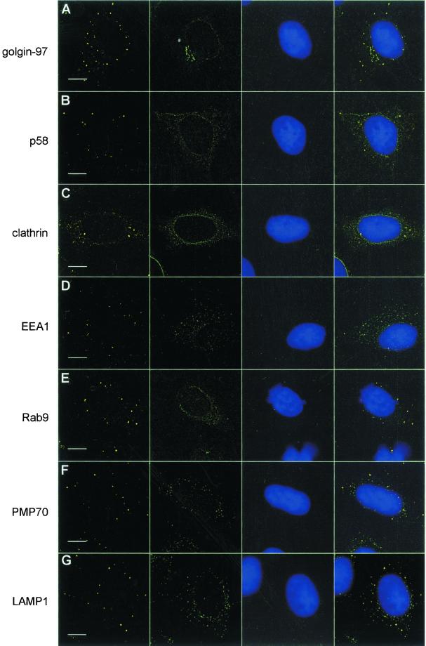
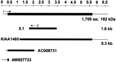
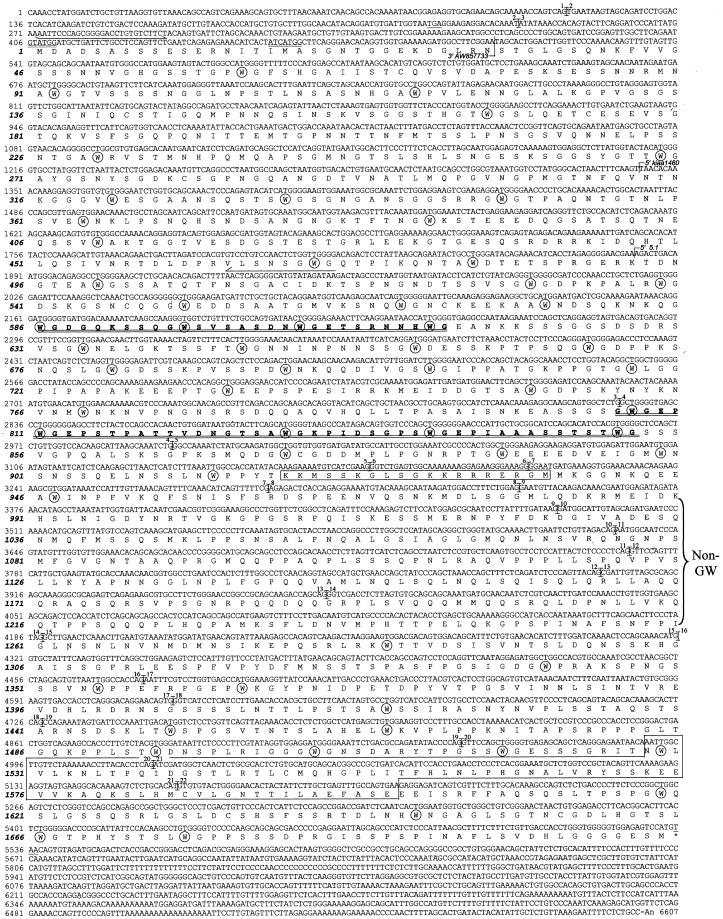
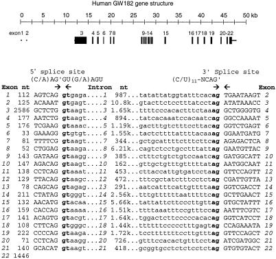
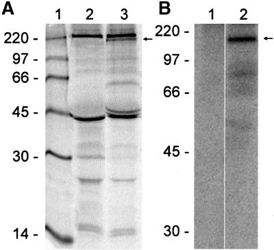
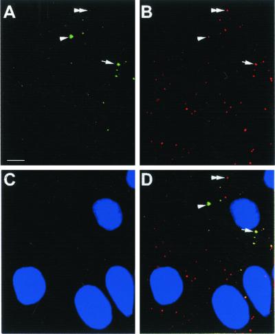
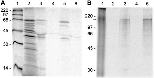
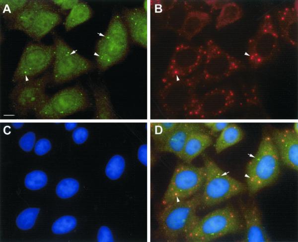
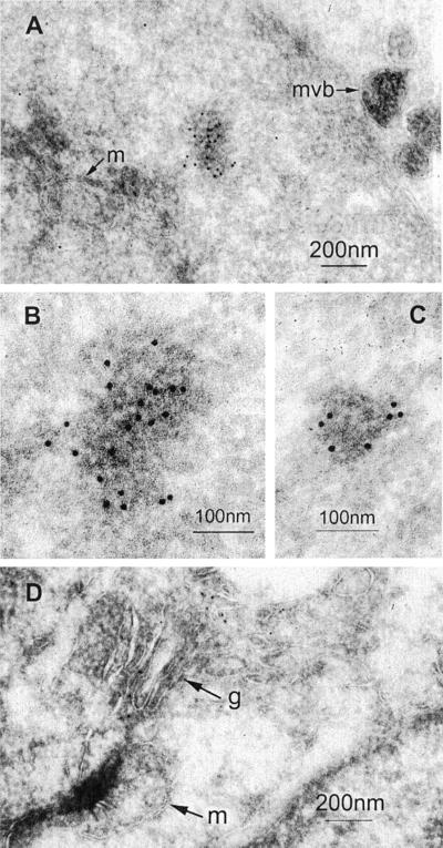
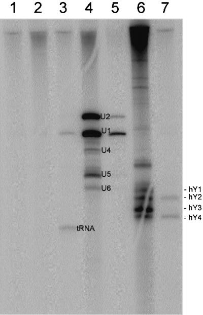
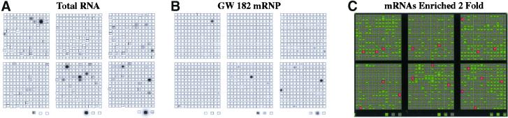
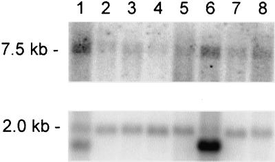
A novel human cellular structure has been identified that contains a unique autoimmune antigen and multiple messenger RNAs. This complex was discovered using an autoimmune serum from a patient with motor and sensory neuropathy and contains a protein of 182 kDa. The gene and cDNA encoding the protein indicated an open reading frame with glycine-tryptophan (GW) repeats and a single RNA recognition motif. Both the patient's serum and a rabbit serum raised against the recombinant GW protein costained discrete cytoplasmic speckles designated as GW bodies (GWBs) that do not overlap with the Golgi complex, endosomes, lysosomes, or peroxisomes. The mRNAs associated with GW182 represent a clustered set of transcripts that are presumed to reside within the GW complexes. We propose that the GW ribonucleoprotein complex is involved in the posttranscriptional regulation of gene expression by sequestering a specific subset of gene transcripts involved in cell growth and homeostasis.


Figures

Figure 1
Figure 1
GW bodies are distinct from known cytoplasmic compartments in HEp-2 cell. The first column represents staining with the index human serum at 1/600 dilution using as a secondary goat anti-human immunoglobulin conjugated to rhodamine. The second column is composed of several different cytoplasmic markers as follows. (A) Mouse monoclonal antigolgin-97 antibody; (B) mouse monoclonal anti-p58 antibody; (C) mouse monoclonal anticlathrin antibody; (D) rabbit anti-EEA1 antibody; (E) mouse monoclonal anti-rab9 antibody; (F) rabbit anti-PMP70 antibody; (G) mouse monoclonal anti-LAMP1 antibody. All these marker antibodies were used at 1/100 dilution. The third column shows corresponding DAPI stained cell nuclei. The fourth column represents images merged from the three columns to the left. The bar lines in the first column represents 10 μm.
Figure 2
Figure 2
A representation of the 5.1 cDNA derived from expression cloning, the EST clone KIAA1460 (GenBank accession no. AB040893), the overlapping genomic fragment AC008731, and EST AW857733 whose combined sequences represent the predicted full-length clone of the GW182 protein. The upstream region was verified using primers 1 and 2, in the predicted 5′-UTR and in the clone 5.1 using HeLa mRNA. Solid boxes represent open reading frame. The full-length protein is 1709 aa and 182 kDa in size.
Figure 3
Figure 3
Human GW182 cDNA and deduced amino acid sequence. The sequence has been submitted to the GenBank under accession no. AY035864. Potential translation start sites (double underlined) and upstream in-frame stop codon (TGA, underlined) are indicated. The GW182 protein consists of 1709 amino acids with a putative nuclear localization signal (NLS; aa 918–937) and RNA recognition motif (RRM; aa 1528–1600) outlined in boxes, respectively. The GW182 protein is rich in tryptophan residues (W, circled), which are often adjacent to glycine (G). Two regions with repeats of GW or WG in tandem are highlighted in bold and double underlined. The region lacking GW is also bracketed. In addition the primers used to amplify the upstream region of the GW182 are shown. Exon–intron junctions and the exact positions of the cDNA clones are indicated.
Figure 4
Figure 4
The human GW182 gene is located on chromosome 16p12 and has 22 exons.
Figure 5
Figure 5
Immunoprecipitation analysis detected a cellular phosphoprotein of ∼180 kDa specifically recognized by the human index serum. (A) Immunoprecipitation of an extract from [35S]methionine-labeled HeLa cells using normal human serum (lane 2) and the human index serum (lane 3) resolved by SDS-PAGE on a 12.5% gel. Lane 1 contains the 14C-labeled molecular weight standard. B. Immunoprecipitation of an extract of [32P]phosphate-labeled HeLa cells using normal human serum (lane 1) and the index human serum (lanes 2) on 10% gel. The specific signals at ∼180 kDa are indicated by arrow. The nonspecific bands at 220 and 45 kDa detected in both lanes 1 and 2 of A are most likely nonmuscle myosin and actin, respectively.
Figure 6
Figure 6
Transfection of HEp-2 cells with a construct of GFP fused to a GW182 cDNA fragment. The transfected GFP-tagged protein produced a staining pattern in transfected cells (A) highly similar to the GWBs recognized by the index human serum (B). When the images in panels A and B were merged with the DAPI stained nuclei of C, colocalization of the GFP product was observed in the cytoplasmic GWBs (D). Three populations of GWBs can be distinguished by the difference in green to red signal. Some GWBs have apparently equal intensity of green and red signals (arrow), whereas others have more green (arrowhead) or more red signal (double arrowhead). Bar, 10 μm. Original magnification, ×600.
Figure 7
Figure 7
Analysis of in vitro–translated GW182 proteins specifically recognized by the index serum and rabbit antibodies raised to recombinant protein. (A) Immunoprecipitation of translation products derived from clone 5.1. Lane 1 shows the 14C-labeled molecular weight standard. Lane 2 shows the proteins produced from in vitro transcription and translation with the largest polypeptide migrating at ∼66 kDa. Immunoprecipitation of the in vitro–translated protein product was performed using the index human serum (lane 3), rabbit preimmune serum (lane 4), postimmune rabbit serum (lane 5), and normal human serum (lane 6). (B) Immunoprecipitation of translation product derived from the full-length construct (lane 1) with the major protein migrating at ∼180 kDa. The latter was immunoprecipitated by the index human serum (lane 2) and rabbit antiserum (lane 5) but not by a normal human serum control (lane 2) and the rabbit preimmune serum (lane 4).
Figure 8
Figure 8
Immunofluorescence analysis of HeLa cell GWBs shows costaining of rabbit anti-GW182 antibody and the index human serum. (A) HeLa cell staining with rabbit anti-GW182 antibody at 1/100 dilution. (B) Staining with the index human serum at 1/600 dilution. (C) Nuclei counterstained with DAPI. (D) Merged image with GWBs (arrowheads) recognized by both antibodies. Note that the rabbit anti-GW182 serum also shows unrelated staining of centrioles (arrows) that is also evident in preimmune rabbit serum (our unpublished results). Bar, 10 μm. Original magnification, ×600.
Figure 9
Figure 9
Immunogold electron microscopy (IEM) localization of GW182 in HeLa cells. Frozen sections of fixed and gelatin-embedded HeLa cells were incubated with the index human serum or rabbit antigiantin diluted 1/400 and then postimmunolabeled with protein A-gold. Examples of the structures labeled by anti-GW182 are shown in A–C and by antigiantin in D. Antibodies to GW182 labeled an electron dense cytoplasmic body that is not bound by a membrane and is distinct from mitochondria (m) and multivesicular bodies (mvb). (B) High-power view of the electron dense body in A. (C) A high-power view of another representative immunogold labeled cytoplasmic electron dense body. Antibodies to giantin label the lateral aspects of the Golgi membrane stack (g). Scale bars are shown in the lower right.
Figure 10
Figure 10
Immunoprecipitation of RNA from 32P-labeled HeLa cell extracts. IP reactions were performed using 100 μl of the cell extract. Normal human serum (lane 1) and the prototype human serum (lane 2) did not precipitate small RNAs. However, prototype anti-tRNA synthetase (lane 3), anti-Sm serum (lane 4), anti-U1RNP serum (lane 5), anti–SS-A/Ro and anti–SS-B/La serum (lane 6), and anti–SS-A/Ro serum (lane 7) all precipitated expected RNA molecules.
Figure 11
Figure 11
Identification of mRNAs associated with GW182-mRNP complexes using cDNA microarray analysis. As described in MATERIALS AND METHODS, RNA was extracted from immunoprecipitated GW182-mRNPs or total cell lysate and used to make reverse-transcribed radiolabeled probes for Atlas Human 1.2 Arrays containing 1188 singly spotted cDNA segments (CLONTECH). Phosphorimages were scanned and stored as gel files and then analyzed using ATLASIMAGE 1.01 software (CLONTECH). A default external background setting was used in conjunction with a background-based signal threshold to determine gene signal significance. Comparison of cDNA array images was performed using an average of all of the gene signals on the array (global normalization) to normalize the signal intensity between arrays. Messenger RNAs associated with GW182–mRNP complexes were considered significant if they were twofold or greater enriched over total cellular RNA. (A) Total cellular RNA; (B) mRNAs associated with GW182–mRNP complexes; (C) computer-generated overlay comparison of representative arrays A and B. Red bars represent species of mRNAs that were enriched twofold or greater as compared with total cellular RNA (genes listed in Table 2). Green bars indicate mRNAs that were present on one or both of the arrays but were not enriched at least twofold.
Figure 12
Figure 12
Northern blot analysis of GW182 mRNA. The order of polyA+ RNA from human tissues was as follows (lanes 1–8): heart, whole brain, placenta, lung, liver, skeletal muscle, kidney, and pancreas. The membrane was probed with a ClaI/EcoRI fragment from the clone KIAA1460 (top panel) and reprobed using actin β-actin cDNA (bottom panel).

References

    1. Ainger K, Avossa D, Diana AS, Barry C, Barbarese E, Carson JH. Transport and localization elements in myelin basic protein mRNA. J Cell Biol. 1997;138:1077–1087. - PMC - PubMed
    1. Alligent J, Aubert S, Dyke KGH, Solh NE. Staphylocoocus caprae strains carry determinants known to be involved in pathogneicity: a gene encoding an autolysin-binding fibronectin and the icaoperon involved in biofilm formation. Infect Immun. 2001;69:712–718. - PMC - PubMed
    1. Altschul SF, Gish W, Miller W, Myers EW, Lipman DJ. Basic local alignment search tool. J Mol Biol. 1990;215:403–410. - PubMed
    1. Antic D, Keene JD. Messenger ribonucleoprotein complexes containing human ELAV proteins: interactions with cytoskeleton and translational apparatus. J Cell Sci. 1998;111:183–197. - PubMed
    1. Bai C, Sen P, Hofmann K, Ma L, Goebl M, Harper JW, Elledge SJ. SKP1 connects cell cycle regulators to the ubiquitin proteolysis machinery through a novel motif, the F-box. Cell. 1996;86:263–274. - PubMed

Publication types

MeSH terms