. 2002 Apr 15;21(8):1939-47.
doi: 10.1093/emboj/21.8.1939.

Conditional switching of VEGF provides new insights into adult neovascularization and pro-angiogenic therapy

Affiliations

Conditional switching of VEGF provides new insights into adult neovascularization and pro-angiogenic therapy

Yuval Dor et al. EMBO J. .

Abstract

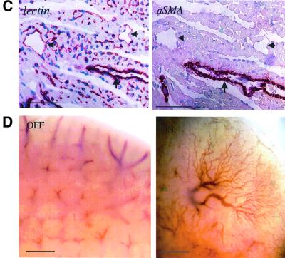
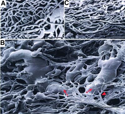
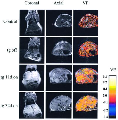
To gain insight into neovascularization of adult organs and to uncover inherent obstacles in vascular endothelial growth factor (VEGF)-based therapeutic angiogenesis, a transgenic system for conditional switching of VEGF expression was devised. The system allows for a reversible induction of VEGF specifically in the heart muscle or liver at any selected schedule, thereby circumventing embryonic lethality due to developmental misexpression of VEGF. Using this system, we demonstrate a progressive, unlimited ramification of the existing vasculature. In the absence of spatial cues, however, abnormal vascular trees were produced, a consequence of chaotic connections with the existing network and formation of irregularly shaped sac-like vessels. VEGF also caused a massive and highly disruptive edema. Importantly, premature cessation of the VEGF stimulus led to regression of most acquired vessels, thus challenging the utility of therapeutic approaches relying on short stimulus duration. A critical transition point was defined beyond which remodeled new vessels persisted for months after withdrawing VEGF, conferring a long-term improvement in organ perfusion. This novel genetic system thus highlights remaining problems in the implementation of pro-angiogenic therapy.


Figures

None
Fig. 1. A transgenic mouse system for conditional switching of VEGF expression in the adult heart or liver. (A) Scheme of the binary transgenic system based on a tetracycline-regulated trans-activator protein driven by either a heart- or liver-specific promoter (‘driver’ lines) and a transgene encoding VEGF164 (‘responder’ line) (see text for details). (B) A heart-specific VEGF switch. VEGF expression was induced by withdrawing tetracycline 1 week after birth and retrieving hearts for analysis 3 weeks later. Upper panels show in situ hybridization with a VEGF probe using myocardial thin sections obtained from littermates control and double transgenic animals that have been processed and hybridized on the same slide. Note induced expression of VEGF in cardiomyocytes. Myocardial hypercellularity observed in the double transgenic heart is due to VEGF-mediated proliferation/recruitment of endothelial cell precursors. Bars, 50 µm. Bottom left: a low magnification view of heart sections hybridized with VEGF as above but autoradiographed through a direct contact with an X-ray film to highlight the overall distribution of VEGF-producing cells. Bottom right: Relative levels of VEGF expression in myocardial sections quantified by autogradiographic grains counts (average ± SD of three high-power fields from each of three independent hybridizations). (C) A liver-specific VEGF switch. VEGF expression was induced by withdrawing tetracycline 1 week after birth and retrieving livers for analysis 4 weeks later. Upper panels show in situ hybridization with a VEGF probe using hepatic thin sections obtained from littermates control and double transgenic animals that have been processed and hybridized on the same slide. Note induced expression of VEGF in hepatocytes. Bars, 50 µm. Bottom left: northern blot analysis of total liver RNA extracted from control animals and from double transgenic animal prior to tetracycline withdrawal (off), 1 month after an ‘on’ switch (on) and 1 month after an ‘on’ switch followed by 3 months in the presence of tetracycline (on>off). Bottom right: Relative levels of VEGF expression were determined by densitometric tracing of the autoradiogram distinguishing the endogenous from transgenic VEGF.
None
Fig. 2. VEGF induces extensive vessel formation in the adult myocardium and liver. (A) Hematoxylin–eosin staining of myocardial sections following a 19-day myocardial VEGF switch. An exaggerated angiogenic response has produced many vessels inter-digitizing muscle fibres and perturbing their normal architecture. Arrows point to the endothelium of these vessels. Bar, 100 µm. (B) A low-power view of the myocardium following a myocardial VEGF switch. Immunostaining with αSMA highlights large coronary vessels covered by VSMCs and pericytes. Note the higher abundance of αSMA-positive vessels in the VEGF-induced myocardium compared with control (quantified as a 2.4-fold increase through counting αSMA-positive vessels in the entire ventricular wall in five sections each). Bar, 1 mm. (C) Right: a higher magnification of the area boxed in (B). Note the presence of both VSMC-coated vessels (arrow) as well as uncoated vessels (arrowheads). Left: immunostaining with an endothelial cell-specific lectin (Bandeiraea simplicifolia BS-1 isolectin) performed on a serial section. Staining appearing in single cells presumably represent sections through capillaries as well as endothelial precursor cells. Bars, 100 µm. (D) Whole-mount views of a liver following a 30-day switch (a bottom view at the edge of the lobes). Note the elaborate vascular tree induced by VEGF. Bar, 1 mm.
None
Fig. 3. Corrosion casts of epicardial vessels. Corrosion casts were prepared and scanned as described in Materials and methods. Data shown are of 10-week-old littermates. (A) Control heart. (B) Four weeks after switching ‘on’ VEGF. Arrows point at examples where flattened vessels merge with hemangioblastoma-like vessels. Arrowhead points at an abnormal multiple branching patterns. (C) Four weeks of ‘on’ followed by 8 days of ‘off’. Bars represent 50 µm in (A) and (C), and 100 µm in (B).
None
Fig. 4. MRI analysis of vascular function and permeability. Left and middle panels: MRI images of the liver. Images shown for a double transgenic animal were derived from the same coronal or axial section taken prior to (tg off) or at the indicated time after switching ‘on’ VEGF. d, days. Note progressive whitening of the image indicative of increased water retention (i.e. edema). Also note the formation of very large vessels discernible even at this low resolution. Right panels: vascular function (VF) in the same axial section shown in the middle panels was determined as described in Materials and methods. A progressive increase in the level of oxygenation is evident, represented on a color-coded relative scale.
None
Fig. 5. Vascular phenotypes resulting from switching off VEGF. (A) Hematoxylin–eosin-stained sections of liver specimens obtained from littermate control (left), tTA+/VEGF+ animals 14 days after switching on VEGF (middle) and from tTA+/VEGF+ animals 14 days after switching on VEGF followed by switching off for 10 days (right). (B) Whole-mount views of livers. tTA+/VEGF+ livers before (left) and after (middle) the indicated times of an ‘on’ switch, and after an additional 5 weeks of an ‘off’ switch (right). Bar, 1 mm. (C) Top view of the entire liver from a control (left) and 4 months after switching off VEGF (following a 4 week stimulus).
None
Fig. 6. Long-term improvement of vascular function in the absence of edema. Monitoring vascular function (A) and edema (B) using MRI was performed as described in the legend to Figure 4. VEGF production was induced in tTA+/VEGF+ animal on day 0 and was switched off either at day 13 (arrow) or at day 32 (arrowhead). Each animal was imaged at the indicated times (squares, short switch; triangles, long switch) and each experimental point represents the average ± SD of readings from four animals. The pre-induction reading in (A) was used as a reference for determining relative increase in liver oxygenation. For monitoring liver edema in (B), the signal intensity in the liver was standardized in each section for the signal intensity in an irrelevant tissue (muscle) whose water content was not affected by VEGF produced by the liver.

References

    1. Abramovitch R., Frenkiel,D. and Neeman,M. (1998) Analysis of subcutaneous angiogenesis by gradient echo magnetic resonance imaging. Magn. Reson. Med., 39, 813–824. - PubMed
    1. Adams R.H. and Klein,R. (2000) Eph receptors and ephrin ligands. Essential mediators of vascular development. Trends Cardiovasc. Med., 10, 183–188. - PubMed
    1. Alon T., Hemo,I., Itin,A., Pe’er,J., Stone,J. and Keshet,E. (1995) Vascular endothelial growth factor acts as a survival factor for newly formed retinal vessels and has implications for retinopathy of prematurity. Nature Med., 1, 1024–1028. - PubMed
    1. Benjamin L.E. and Keshet,E. (1997) Conditional switching of vascular endothelial growth factor (VEGF) expression in tumors: induction of endothelial cell shedding and regression of hemangioblastoma-like vessels by VEGF withdrawal. Proc. Natl Acad. Sci. USA, 94, 8761–8766. - PMC - PubMed
    1. Benjamin L.E., Hemo,I. and Keshet,E. (1998) A plasticity window for blood vessel remodelling is defined by pericyte coverage of the preformed endothelial network and is regulated by PDGF-B and VEGF. Development, 125, 1591–1598. - PubMed

Publication types

Substances