. 2002 Apr 15;21(8):1967-77.
doi: 10.1093/emboj/21.8.1967.

Caspase-6 gene disruption reveals a requirement for lamin A cleavage in apoptotic chromatin condensation

Affiliations

Caspase-6 gene disruption reveals a requirement for lamin A cleavage in apoptotic chromatin condensation

Sandrine Ruchaud et al. EMBO J. .

Abstract

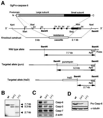
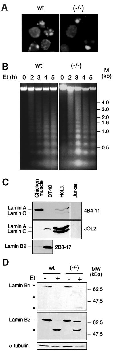
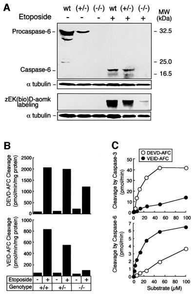
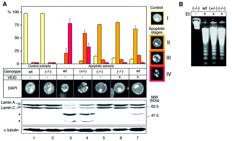
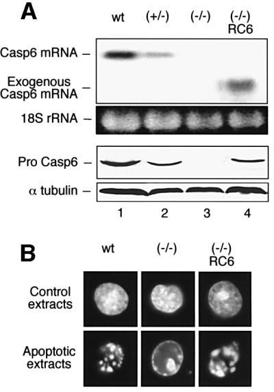
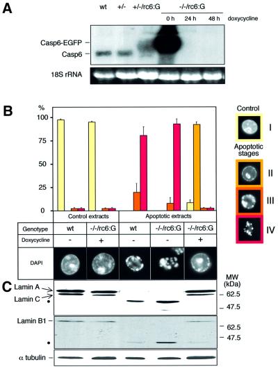
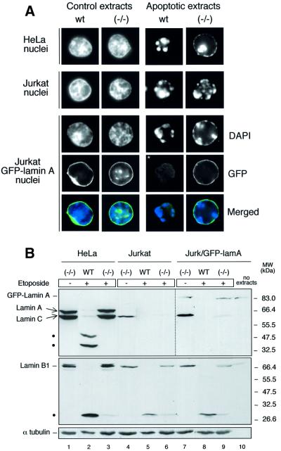
To study the role of caspase-6 during nuclear disassembly, we generated a chicken DT40 cell line in which both alleles of the caspase-6 gene were disrupted. No obvious morphological differences were observed in the apoptotic process in caspase-6- deficient cells compared with the wild type. However, examination of apoptosis in a cell-free system revealed a block in chromatin condensation and apoptotic body formation when nuclei from HeLa cells expressing lamin A or lamin A-transfected Jurkat cells were incubated in caspase-6-deficient apoptotic extracts. Transfection of exogenous caspase-6 into the clone reversed this phenotype. Lamins A and C, which are caspase-6-only substrates, were cleaved by the wild-type and heterozygous apoptotic extracts but not by the extracts lacking caspase-6. Furthermore, the caspase-6 inhibitor z-VEID-fmk mimicked the effects of caspase-6 deficiency and prevented the cleavage of lamin A. Taken together, these observations indicate that caspase-6 activity is essential for lamin A cleavage and that when lamin A is present it must be cleaved in order for the chromosomal DNA to undergo complete condensation during apoptotic execution.


Figures

None
Fig. 1. Structure and targeting of the Gallus gallus caspase-6 gene. (A) Structure of the chicken caspase-6 gene together with the targeting vectors and homologous recombinants containing either the puromycin or the histidinol cassette. (B–D) Analysis of the caspase-6 homozygous (wt), heterozygous (+/–) and null clones (–/–). (B) Southern blot analysis of DNA digested by BamHI, using the 3′ genomic external probe ApaI–BamHI represented in (A). (C) Northern blot analysis showing chicken caspase-6, -3 and -7 mRNA expression. Note the loss of caspase-6 mRNA in the null clone. (D) Immunoblotting of caspase-6 using a polyclonal antibody directed against the large subunit of the enzyme (R549).
None
Fig. 2. Apoptosis in caspase-6-deficient DT40 cells is phenotypically normal. (A) DAPI staining of wild-type and caspase-6–/– cells treated with 10 µM etoposide for 4 h. Apoptosis was quantified by TUNEL labelling in this experiment, showing 58% apoptotic cells in the wild-type and 35% in the caspase-6–/– cells; no difference was observed when cells were treated with staurosporine (data not shown). (B) DNA fragmentation during the etoposide time course on wild-type and caspase-6–/– cells. (C) Immunoblotting analysis of DT40, Jurkat and HeLa whole-cell lysates together with chicken muscle tissue lysate, using monoclonal antibodies to human lamin A/C (JOL2), chicken lamin A (4B4-11) or chicken lamin B2 (2B8-17). This last antibody does not recognize the human form of lamin B2. The signal in the right lanes (Jurkat) was enhanced relative to that in the left lanes. (D) Immunoblotting analysis of wild-type DT40 and caspase-6–/– cells treated or not by etoposide at 10 µM for 4 h, using monoclonal antibodies to chicken lamin B1 and B2 (clone L-5 and 2B8-17, respectively). The cleavage products are shown by black dots.
None
Fig. 3. Characterization of the in vitro apoptosis system and apoptotic extracts from DT40 cells. (A) Caspase-6 expression analysis by immunoblotting in extracts from wild-type, (+/–) and (–/–) cells treated with 10 µM etoposide or diluent for 5 h. When caspase-6 in the apoptotic extracts is processed, the antibody (R549) recognizes the large subunit at ∼20 kDa. The lower panel shows the labelling of active caspases by zEK(bio)D-aomk. α-tubulin expression is used as gel loading control. (B) Measurement of DEVD-AFC and VEID-AFC cleavage activity in cytosol from etoposide-treated DT40 cells. Similar results were obtained when cytosol was prepared from staurosporine-treated DT40 clones (data not shown). (C) Evaluation of the selectivity of recom binant caspases-3 and -6 toward DEVD-AFC and VEID-AFC. Note the substantial cleavage of VEID-AFC by caspase-3 and DEVD-AFC by caspase-6 under widely used reaction conditions despite the relatively low affinity of the enzymes for these non-preferred substrates.
None
Fig. 4. A block in apoptotic chromatin condensation in caspase-6-deficient apoptotic extracts. Isolated HeLa nuclei were incubated for 2 h in the apoptotic extracts previously characterized from wild-type, (+/–) and (–/–) cells (see Figure 3) in the presence (+) or absence (–) of the caspase-6-specific inhibitor VEID-fmk at 1 µM. (A) For each extract, the number of nuclei in apoptotic stages (I–IV) was determined. The stages were defined following DAPI staining of HeLa nuclei incubated in etoposide-treated DT40 apoptotic extracts or control DT40 extracts. HeLa nuclei incubated in control extracts define stage I. After addition to apoptotic extracts, the chromatin begins to condense against the nuclear periphery and nucleoli (stage II). Next, the peripheral chromatin ring condenses into discrete masses that separate from one another while the condensed chromatin from the nucleolus migrates to the nuclear periphery (stage III). Finally, the chromatin masses form discrete apoptotic bodies and the nuclear shape is lost (stage IV). A nucleus representative of the major population is shown for each stage. Data shown represent three independent experiments with an average of 300 nuclei counted per condition. After the 2 h incubation in the extracts, lamin A/C cleavage from HeLa nuclei was assessed by immunoblotting using the monoclonal antibody to human lamin A/C, JOL2. The cleavage products are shown by black dots. α-tubulin expression is used as gel loading control. Controls in this experiment include extracts from untreated wild-type cells, which gave 98% of uncondensed (non-apoptotic) nuclei upon incubation. The low (2%) frequency of apoptotic nuclei seen in controls reflects apoptotic cells in the HeLa population used to prepare nuclei, as this low percentage is always observed even at early incubation times. (B) Analysis of DNA fragmentation in the HeLa nuclei after the 2 h incubation in the extracts.
None
Fig. 5. The knockout phenotype is rescued by exogenous caspase-6. The caspase-6–/– cells were transfected with a construct containing the chicken caspase-6 cDNA under control of a CMV promoter. (A) Analysis of the exogenous caspase-6 expression in one transfected clone RC6. Upper panels: mRNA expression analysed by northern blotting. Lower panels: protein expression analysed by immunoblotting using polyclonal anti-caspase-6 (R549). (BIn vitro changes induced in HeLa nuclei by control and apoptotic extracts from wild-type, caspase-6–/– and caspase-6–/–/RC6 cells.
None
Fig. 6. The knockout phenotype is observed in an independent caspase-6-deficient clone. Analysis of an independent knockout clone (designated caspase-6–/–/casp-6:EGFP in the text and –/–/rc6:G in the figure) expressing the caspase-6:EGFP fusion protein under the control of the tetracycline-repressible system. (A) mRNA expression of endogenous caspase-6 and caspase-6:EGFP in the wild-type, +/–, +/–/rc6:G clones and –/–/rc6:G clone grown in the absence or presence of 1 µg/ml doxycycline for 24 and 48 h. (B) Isolated HeLa nuclei were incubated for 2 h in apoptotic extracts from wild-type cells and from –/–/rc6:G cells grown in the absence or presence of 1 µg/ml doxycycline for 2 weeks prior to induction of apoptosis. Apoptotic nuclei were counted according to the procedure described in Figure 4A. (C) After the 2 h incubation in the extracts, lamin A/C and B1 integrity in HeLa nuclei was assessed by immunoblotting using the antibody to lamin A/C (JOL2) and the antibody to lamin B1 (clone L-5). α-tubulin is used as gel loading control. The cleavage products are indicated by black dots.
None
Fig. 7. Lamin A cleavage by caspase-6 is required for chromatin condensation and nuclear disassembly. (A) Isolated HeLa, Jurkat or Jurkat:GFP–lamin A nuclei were incubated for 2 h in extracts from wild-type and caspase-6–/– cells treated with 1 µM staurosporine or diluent for 8 h. A nucleus representative of the major population is shown for each condition (DAPI) along with the GFP in the Jurkat:GFP–lamin A nuclei. The signal in the panel indicated by a white asterisk was enhanced relative to that in the other panels in order to see the residual GFP–lamin A fluorescence. (B) After the 2 h incubation in the extracts, the integrity of lamin A/C and B1 from HeLa nuclei was assessed by immunoblotting using the antibody to lamin A/C (JOL2) and the antibody to lamin B1 (clone L-5). The cleavage products are indicated by black dots. Non-adjacent wells on the same blot have been juxtaposed to compose each panel in this figure. The signal in lanes 7–10 was enhanced relative to that in the other panels in order to see the GFP–lamin A band.

References

    1. Allsopp T.E., McLuckie,J., Kerr,L.E., Macleod,M., Sharkey,J. and Kelly,J.S. (2000) Caspase 6 activity initiates caspase 3 activation in cerebellar granule cell apoptosis. Cell Death Differ., 7, 984–993. - PubMed
    1. Benavente R., Krohne,G. and Franke,W.W. (1985) Cell type-specific expression of nuclear lamina proteins during development of Xenopus laevis. Cell, 41, 177–190. - PubMed
    1. Bonne G. et al. (1999) Mutations in the gene encoding lamin A/C cause autosomal dominant Emery–Dreifuss muscular dystrophy. Nature Genet., 21, 285–288. - PubMed
    1. Budihardjo I., Oliver,H., Lutter,M., Luo,X. and Wang,X. (1999) Biochemical pathways of caspase activation during apoptosis. Annu. Rev. Cell Dev. Biol., 15, 269–290. - PubMed
    1. Buerstedde J.-M. and Takeda,S. (1991) Increased ratio of targeted to random integration after transfection of chicken B cell lines. Cell, 67, 179–188. - PubMed

Publication types