. 2002 Apr 15;21(8):2038-44.
doi: 10.1093/emboj/21.8.2038.

Specific interaction of IP6 with human Ku70/80, the DNA-binding subunit of DNA-PK

Affiliations

Specific interaction of IP6 with human Ku70/80, the DNA-binding subunit of DNA-PK

Les A Hanakahi et al. EMBO J. .

Abstract

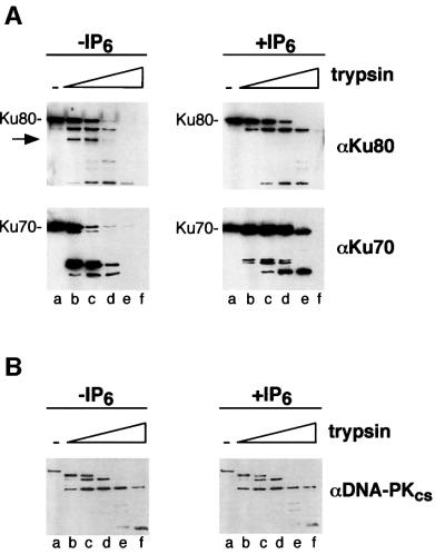
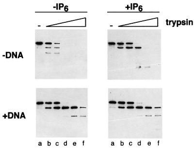
In eukaryotic cells, DNA double-strand breaks can be repaired by non-homologous end-joining, a process dependent upon Ku70/80, XRCC4 and DNA ligase IV. In mammals, this process also requires DNA-PK(cs), the catalytic subunit of the DNA-dependent protein kinase DNA-PK. Previously, inositol hexakisphosphate (IP6) was shown to be bound by DNA-PK and to stimulate DNA-PK-dependent end-joining in vitro. Here, we localize IP6 binding to the Ku70/80 subunits of DNA- PK, and show that DNA-PK(cs) alone exhibits no detectable affinity for IP6. Moreover, proteolysis mapping of Ku70/80 in the presence and absence of IP6 indicates that binding alters the conformation of the Ku70/80 heterodimer. The yeast homologue of Ku70/80, yKu70/80, fails to bind IP6, indicating that the function of IP6 in non-homologous end-joining, like that of DNA-PK(cs), is unique to the mammalian end-joining process.


Figures

None
Fig. 1. Specific binding of IP6 by purified DNA-PK. Binding reactions contained 5000 U of purified DNA-PK (Promega) and 100 nM [3H]IP6, in the presence or absence of unlabelled competitor as indicated. Complexes were separated by gel filtration through Superdex 200. [3H]IP6 was detected by scintillation counting. (A) DNA-PK with [3H]IP6 only. (B) As (A), but in the presence of a 100-fold excess of IS6. (C) As (A), but in the presence of a 10-fold excess of IP6. (D) Control indicating the mobility of [3H]IP6 in the presence of non-specific marker proteins.
None
Fig. 2. Interaction of IP6 with components of the DNA-PK holoenzyme. Ku70/80 (300 nM), DNA-PKcs (300 nM) and [3H]IP6 (100 nM) were mixed and incubated as described in Materials and methods. The products were then resolved by gel filtration through Superdex 200. [3H]IP6 was detected by scintillation counting; Ku70/80 and DNA-PKcs were detected by western blotting. (A) Binding of IP6 by DNA-PK. (B) Reactions were carried out as described for (A), but in the presence of dsDNA (300 nM).
None
Fig. 3. Specific interaction of IP6 with Ku70/80. Spin-column analysis of the binding of [3H]IP6 by Ku70/80. Following centrifugation, the [3H]IP6 present in the void volume indicates the amount bound by Ku70/80. (A) Binding reactions were carried out as described in Materials and methods using [3H]IP6 (100 nM) and Ku70/80 (filled squares) or DNA-PKcs (open diamonds). (B) Binding reactions were carried out with Ku70/80 (100 nM) and [3H]IP6 (100 nM), and supplemented with unlabelled IP6 (1, 3 and 10 times molar excess) or unlabelled IS6 (10 times molar excess).
None
Fig. 4. Trypsin proteolysis mapping of Ku70/80 and DNA-PKcs in the presence and absence of IP6. (A) Trypsin digestion of Ku70/80. Protein (1.3 µg) was digested in the presence or absence of IP6 (10 µM) using the following amounts of trypsin: 0, 1.6, 4, 16, 44 and 132 ng (lanes a– f). The arrow shows a proteolysis product that is formed only in the absence of IP6. (B) Trypsin digestion of DNA-PKcs. Protein (1.3 µg) was digested with trypsin as described in (A). Proteins were detected by western blotting.
None
Fig. 5. V8 protease mapping of Ku70/80 in the presence and absence of IP6. Reactions were carried out as described in Figure 4, using the following amounts of V8 protease: 0, 4, 16, 48, 148 and 444 ng (lanes a–f). In the lower panels, a slight cross-reactivity of the anti-Ku70 polyclonal antiserum to Ku80 can be seen.
None
Fig. 6. Trypsin proteolysis mapping of Ku70/80–DNA complexes. Reactions were carried out as described in the legend to Figure 4, using Ku70/80 in the presence or absence of sheared calf thymus DNA. Proteins were detected with western blotting using anti-Ku80 poly clonal antibodies.
None
Fig. 7. Specificity of IP6 for mammalian Ku70/80. Spin-column analysis of the binding of [3H]IP6 by Ku70/80. Following centrifugation, the [3H]IP6 present in the void volume indicates the amount bound by Ku70/80. Error bars represent the standard deviation of three independent measurements. (A) Comparison of the binding of human Ku70/80 (100 nM) or its yeast homologue yKu70/80 (1 µM) with [3H]IP6 (100 nM). (B) Comparison of the [3H]IP6 (100 nM) binding ability of purified recombinant Ku70 (500 nM), recombinant Ku80 (500 nM), recombinant Ku70/80 containing a C-terminal truncated Ku80 subunit [rKu70/80(T); 100 nM], recombinant Ku70/80 containing a full-length Ku80 subunit [rKu70/80(FL); 100 nM], and Ku70/80 purified from HeLa cells [Ku70/80 (HeLa); 100 nM].

References

    1. Bailey S.M., Meyne,J., Chen,D.J., Kurimasa,A., Li,G.C., Lehnert,B.E. and Goodwin,E.H. (1999) DNA double-strand break repair proteins are required to cap the ends of mammalian chromosomes. Proc. Natl Acad. Sci. USA, 96, 14899–14904. - PMC - PubMed
    1. Chan D.W. and Lees-Miller,S.P. (1996) The DNA-dependent protein kinase is inactivated by autophosphorylation of the catalytic subunit. J. Biol. Chem., 271, 8936–8941. - PubMed
    1. Chan D.W., Ye,R.Q., Veillette,C.J. and Lees-Miller,S.P. (1999) DNA-dependent protein kinase phosphorylation sites in Ku70/80 heterodimer. Biochemistry, 38, 1819–1828. - PubMed
    1. Chu G. (1997) Double-strand break repair. J. Biol. Chem., 272, 24097–24100. - PubMed
    1. Critchlow S.E. and Jackson,S.P. (1998) DNA end-joining: from yeast to man. Trends Biochem. Sci., 23, 394–398. - PubMed

Publication types