. 2002 Apr 15;21(8):2045-53.
doi: 10.1093/emboj/21.8.2045.

The active site of the DNA repair endonuclease XPF-ERCC1 forms a highly conserved nuclease motif

Affiliations

The active site of the DNA repair endonuclease XPF-ERCC1 forms a highly conserved nuclease motif

Jacqueline H Enzlin et al. EMBO J. .

Abstract

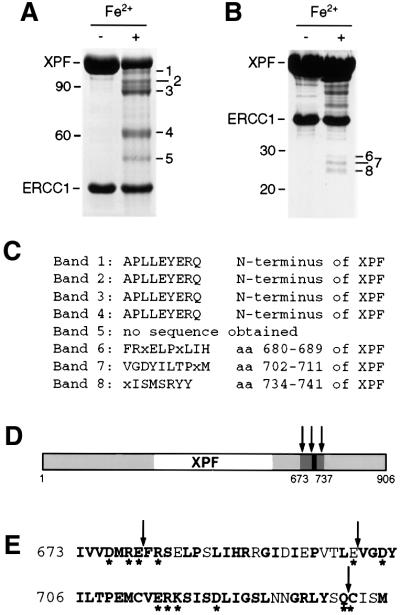
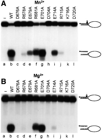
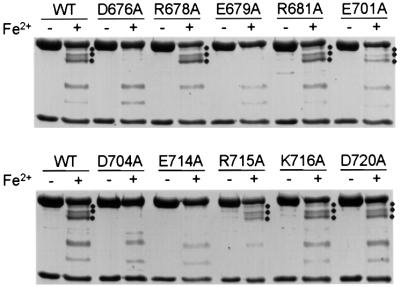
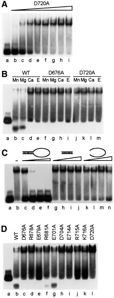
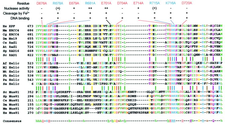
XPF-ERCC1 is a structure-specific endonuclease involved in nucleotide excision repair, interstrand crosslink repair and homologous recombination. So far, it has not been shown experimentally which subunit of the heterodimer harbors the nuclease activity and which amino acids contribute to catalysis. We used an affinity cleavage assay and located the active site to amino acids 670-740 of XPF. Point mutations generated in this region were analyzed for their role in nuclease activity, metal coordination and DNA binding. Several acidic and basic residues turned out to be required for nuclease activity, but not DNA binding. The separation of substrate binding and catalysis by XPF-ERCC1 will be invaluable in studying the role of this protein in various DNA repair processes. Alignment of the active site region of XPF with proteins belonging to the Mus81 family and a putative archaeal RNA helicase family reveals that seven of the residues of XPF involved in nuclease activity are absolutely conserved in the three protein families, indicating that they share a common nuclease motif.


Figures

None
Fig. 1. Purification of recombinant XPF–ERCC1. (A) Comparison of the gel-filtration profiles of XPF–ERCC1 expressed in E.coli (dotted line) and Sf9 insect cells (solid line). The peak at ∼45 ml corresponds to the void volume of the column with protein in an aggregated state, the peak at ∼65 ml to a molecular weight of ∼200 kDa and the peak at ∼75 ml to a molecular weight of ∼60 kDa. The proteins identified in the peak fractions are indicated. (B) SDS–PAGE gel (10%) of the purification of the XPF–ERCC1 heterodimer from Sf9 cells; proteins were visualized by CBB staining. The cell lysate (Lys) and pooled fractions from after the individual column steps, Ni-NTA–agarose (Ni), gel filtration (Gf) and heparin (Hep), are shown. XPF and ERCC1 have molecular weights of 115 and 40 kDa, respectively.
None
Fig. 2. Iron-induced cleavage of XPF–ERCC1. (A) SDS–PAGE gel (8%) of purified XPF–ERCC1 before and after treatment with 50 µM FeCl2 and 20 mM ascorbate for 15 h at 0°C. The five observed XPF cleavage products were designated bands 1–5, in order of decreasing size and with estimated molecular weights of 102, 95, 88, 62 and 52 kDa, respectively. A 10 kDa protein ladder (Life Technologies) was used as a marker. The positions of the 90 and 60 kDa marker bands are indicated. (B) SDS–PAGE gel (12%) of XPF–ERCC1 treated as in (A). The three bands between 25 and 30 kDa were designated bands 6–8. The positions of the 30 and 20 kDa marker bands are indicated. (C) Results from the N-terminal sequencing of the peptide fragments from bands 1–8 in (A) and (B). (D) Location of the cleavage sites in XPF. Regions of XPF that are conserved between species are shown in gray, the region encompassing the cleavage sites in dark gray and the highly conserved V/IERKX3D motif in black. The cleavage sites are indicated by arrows. (E) The amino acid sequence of region 673–737 of XPF. Conserved residues are in bold, residues selected for site- directed mutagenesis are marked with an asterisk and cleavage sites are marked with arrows.
None
Fig. 3. Nuclease activity of wild-type and mutant XPF–ERCC1. Incision of a stem12–loop22 DNA substrate (100 fmol) by wild-type (WT, lane b) or mutant (lanes c–l) XPF–ERCC1 (200 fmol) in the presence of (A) 0.4 mM MnCl2 or (B) 2 mM MgCl2. Reactions were analyzed on a 15% denaturing polyacrylamide gel. The XPF mutations tested, the 46mer stem–loop substrate (with incision sites indicated by arrows) and the 9/10mer products resulting from cleavage are indicated.
None
Fig. 4. Comparison of iron-induced cleavage of wild-type and mutant XPF–ERCC1. Purified wild-type (WT) and mutant XPF–ERCC1 were incubated with 50 µM FeCl2 and 20 mM ascorbate for 15 h at 0°C and analyzed on an 8% SDS–PAGE gel. Bands 1–3 resulting from iron- induced cleavage, with molecular weights of ∼102, 95 and 88 kDa, respectively, are indicated (where present) by three dots.
None
Fig. 5. Catalytically inactive XPF–ERCC1 mutants bind specifically to stem–loop substrates. 5′-32P-labeled stem12–loop22 substrate at a concentration of 17 nM was used for all experiments and was the last component added to the reaction mixture. Unless indicated otherwise, a protein concentration of 68 nM and MnCl2 (0.4 mM) as a cofactor were used. Reactions were incubated for 30 min at room temperature and analyzed on 4% native polyacrylamide gels. (A) Increasing concentrations of XPF(D720A)–ERCC1 were incubated with the substrate to demonstrate binding of XPF–ERCC1 to the stem–loop substrate. Protein concentrations: lane b, 17 nM; lane c, 34 nM; lane d, 51 nM; lane e, 68 nM; lane f, 85 nM; lane g, 102 nM; lane h, 136 nM; lane i, 170 nM. (B) Influence of the presence of divalent metal ions on substrate binding by wild-type and mutant XPF–ERCC1. Wild-type (WT) XPF–ERCC1, lanes b–e; D676A, lanes f–i; D720A, lanes j–m. Concentrations of metal cofactors: Mn2+, 0.4 mM; Mg2+, 2 mM; Ca2+, 2 mM. Concentration of EDTA (E), 5 mM. (C) Influence of increasing amounts of specific and non-specific oligonucleotide competitors on substrate binding by XPF(D720A)–ERCC1. Competitors: lane b, no competitor; lanes c–f, stem12–loop22 (68, 340, 850 and 1700 nM, respectively); lanes g–j, stem12 dsDNA (340, 850, 1700 and 3400 nM, respectively); lanes k–n, loop22 ssDNA (340, 850, 1700 and 3400 nM, respectively). (D) Comparison of substrate binding of wild-type (WT) and mutant XPF–ERCC1.
None
Fig. 6. Alignment of the endonuclease domain of XPF with its homologs. The alignment shows amino acids 673–737 of human XPF with homologous proteins of the XPF, putative archaeal helicase and Mus81 family. The aligned proteins (DDBJ/EMBL/GenBank accession Nos are shown in parentheses): human (Hs) XPF (Q92889), mouse (Mm) ERCC4 (AAC03240), hamster (Cg) ERCC4 (BAA89299), Drosophila melanogaster (Dm) Mei9 (Q24087), Caenorhabditis elegans (Ce) Rad1 (CAB70217), Arabidopsis thaliana (At) Rad1 (NP_198931), Schizosaccharomyces pombe (Sp) RAD16 (P36617), Saccharomyces cerevisiae (Sc) RAD1 (P06777), Archaeoglobus fulgidus (Af) putative RNA helicase (AAB89786), Methanococcus jannaschii (Mj) putative RNA helicase (AAB99518), Methanothermobacter thermautotrophicus (Mt) putative RNA helicase (AAB85892), Pyrococcus horikoshii (Ph) putative RNA helicase (NP_143722), Pyrococcus abyssi (Pa) putative RNA helicase (NP_125972), Sulfolobus solfataricus (Ss) putative RNA helicase (CAB57574), Sc Mus81 (NP_010674), Sp Mus81 (CAB09772), Ce Mus81 (AAB37627), Dm Mus81 (AAF45571), Mm Mus81 (AAL28066), Hs Mus81 (AAL28065). Conserved residues are indicated in the following colors: green, hydrophobic (I,L,V,M); red, acidic (D,E); blue, basic (K,R,H); brown, aromatic (F,Y,W); yellow (G,P); purple (C,N,Q,S,T); orange (A). Residues that are ≥85% conserved are indicated in the consensus sequence; absolutely conserved residues are underlined. Conserved hydrophobic (h) and aromatic (a) residues are indicated. Results from the biochemical characterization of the mutants generated in this study are summarized above the alignment.

References

    1. Aboussekhra A. et al. (1995) Mammalian DNA nucleotide excision repair reconstituted with purified protein components. Cell, 80, 859–868. - PubMed
    1. Araujo S.J., Nigg,E.A. and Wood,R.D. (2001) Strong functional interactions of TFIIH with XPC and XPG in human DNA nucleotide excision repair, without a preassembled repairosome. Mol. Cell. Biol., 21, 2281–2291. - PMC - PubMed
    1. Aravind L., Walker,D.R. and Koonin,E.V. (1999) Conserved domains in DNA repair proteins and evolution of repair systems. Nucleic Acids Res., 27, 1223–1242. - PMC - PubMed
    1. Bardwell A.J., Bardwell,L., Tomkinson,A.E. and Friedberg,E.C. (1994) Specific cleavage of model recombination and repair intermediates by the yeast Rad1–Rad10 DNA endonuclease. Science, 265, 2082–2085. - PubMed
    1. Biggerstaff M., Szymkowski,D.E. and Wood,R.D. (1993) Co-correction of the ERCC1, ERCC4 and xeroderma pigmentosum group F DNA repair defects in vitro. EMBO J., 12, 3685–3692. - PMC - PubMed

Publication types

MeSH terms