. 2002 May;70(5):2566-75.
doi: 10.1128/IAI.70.5.2566-2575.2002.

Glucan is a component of the Mycobacterium tuberculosis surface that is expressed in vitro and in vivo

Affiliations

Glucan is a component of the Mycobacterium tuberculosis surface that is expressed in vitro and in vivo

J Reid Schwebach et al. Infect Immun. 2002 May.

Abstract

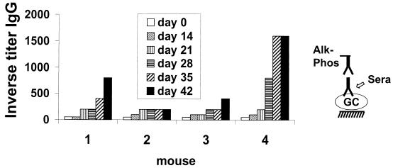
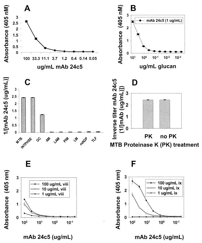
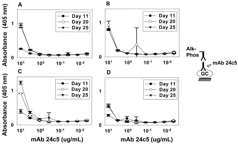
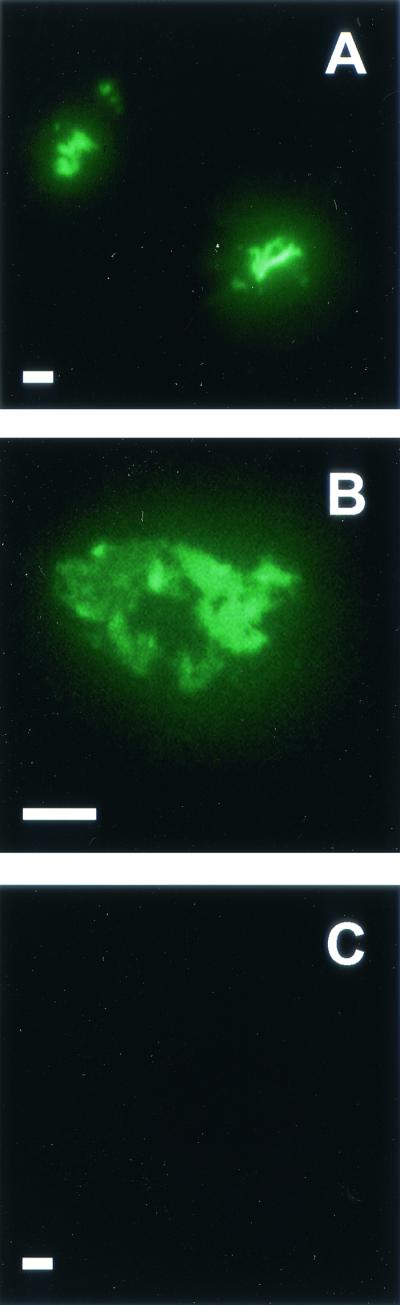
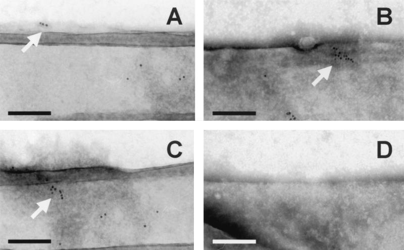
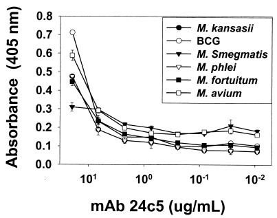
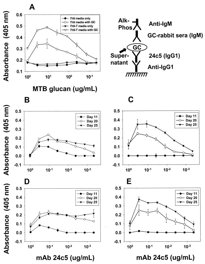
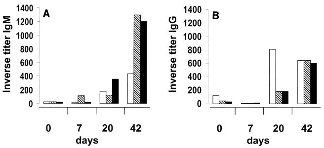
The outermost layer of Mycobacterium tuberculosis is composed primarily of two polysaccharides, glucan (GC) and arabinomannan. To analyze the surface polysaccharide composition of M. tuberculosis, we generated a monoclonal antibody (MAb) that binds M. tuberculosis GC and is known as MAb 24c5. Immunofluorescence and whole-mount immunoelectron microscopy indicated that GC is on the outermost portion of the bacteria. M. tuberculosis strains Erdman and CDC 1551 were analyzed for their ability to bind MAb 24c5 after in vitro growth in media with and without the detergent Tween 80. MAb 24c5 bound to Erdman and CDC 1551 at all culture times with only slightly greater apparent affinity after extended culture in the absence of Tween 80, indicating that a stable amount of GC polysaccharide antigen is associated with the cell surface of M. tuberculosis. An enzyme-linked immunosorbent assay indicated that GC is antigenically similar to glycogen, and the amount of GC antigen increased in the media of M. tuberculosis cultures grown either with or without the detergent Tween 80. Other nontuberculosis mycobacteria have antigenically similar GCs on their surfaces after in vitro growth. Inoculation of mice with live bacilli but not inoculation with dead bacilli elicited a strong antibody response to GC consistent with production of this antigen in vivo. Our results provide a more comprehensive picture of the M. tuberculosis cell envelope and the conditions that allow expression of M. tuberculosis GC.


Figures

FIG. 1.
FIG. 1.
Immunogenicity of GC-rEPA: anti-GC IgG titers of four different mice after immunization and boosting with GC-rEPA. A diagram of the ELISA used to determine titers is on the right. Alk-Phos, alkaline phosphatase.
FIG. 2.
FIG. 2.
MAb 24c5 ELISA analysis and binding to glycogen. (A) Splenic fusion resulted in MAb 24c5, an IgG1 able to recognize M. tuberculosis GC. Each data point represents the average of three measurements. (B) MAb 24c5 binding to dilute concentrations of GC in the M. tuberculosis GC ELISA. Each data point represents the average of three measurements. (C) MAb 24c5 binding to M. tuberculosis Erdman day-20 7H9 medium (MTB), cell culture supernatant extract (INTPHSE), GC, AM, lipoarabinomannan (LAM), phosphatidylinositol mannoside (PIM), fast-growing mycobacterial lipomannan (LM), mycolyl-arabinogalactan-peptidoglycan complex (mAGP), and total lipid fraction (TLF). Each bar represents the average of two measurements. (D) Effects of proteinase K on binding of MAb 24c5 to M. tuberculosis Erdman day-20 7H9 medium. (E and F) MAb 24c5 assayed for reactivity to type VIII (slipper limpet) (viii) (E) and type IX (ix) (bovine liver) (F) glycogens. Each data point represents the average of three measurements. Error bars show the standard deviations of the means.
FIG. 3.
FIG. 3.
Binding of MAb 24c5 to whole cells of M. tuberculosis Erdman and CDC 1551 at various times during culture. M. tuberculosis Erdman (A and B) and CDC 1551 (C and D) were grown in the absence (A and C) or in the presence (B and D) of Tween 80 for 11, 20, or 25 days before harvest. Each data point represents the average of three measurements. Error bars show the standard deviations of the means. The growth experiments and the ELISA analysis were repeated twice, with similar results. Alk-Phos, alkaline phosphatase.
FIG. 4.
FIG. 4.
IF microscopy of M. tuberculosis Erdman using MAb 24c5. Formalin-fixed M. tuberculosis Erdman grown in the absence of Tween for 25 days was analyzed by IF microscopy using ALEXA-488-conjugated MAb 24c5 (A and B) or irrelevant control MAb (C). Bars = 5 μm. Slides were viewed at a magnification of ×100 using oil immersion. For photography an exposure time of 27 s was used for all samples. Slides were scanned into Adobe Photoshop, version 5.0, and images were transferred to Microsoft Powerpoint, version 2000, without any modification except enlargement.
FIG. 5.
FIG. 5.
Immunoelectron microscopy of M. tuberculosis using MAb 24c5. M. tuberculosis Erdman grown in the absence of Tween 80 for 25 days was analyzed for binding of MAb 24c5 (A to C) by using goat anti-mouse IgG sera conjugated to 5-nm gold. Gold appears as black dots, as indicated by arrows (A to C). The images in panels A to C are from three different bacterial fields and two different M. tuberculosis samples. Irrelevant control antibody did not allow binding of gold-conjugated sera (D). Bars = 200 nm.
FIG. 6.
FIG. 6.
Binding of MAb 24c5 to other mycobacteria. Mycobacteria were grown in Sauton's medium without Tween 80 before harvest. Equal volumes of mycobacteria were used in ELISA. Each data point represents the average of three separate measurements. Error bars show the standard deviations of the means.
FIG. 7.
FIG. 7.
GC capture ELISA and analysis of M. tuberculosis culture supernatants. The diagram shows the capture ELISA employing MAb 24c5 (Alk-Phos, alkaline phosphatase). This assay was able to detect the presence of a standard amount of M. tuberculosis (MTB) GC added to culture supernatant with or without the detergent Tween 80 (A). (B to E) M. tuberculosis Erdman (B and C) and CDC 1551 (D and E) were grown in the absence (B and D) or in the presence (C and E) of Tween 80 for 11, 20, or 25 days before the supernatant was harvested and the capture ELISA was performed. The binding to medium alone was subtracted from the values shown in panels B to E. Each data point represents the average of two measurements. Error bars show the standard deviations of the means.
FIG. 8.
FIG. 8.
Antibody titer to GC during M. tuberculosis infection. Murine serum was assayed for IgM (A) and IgG (B) antibody titers to GC at zero time and 7, 20, and 42 days after infection of mice with M. tuberculosis Erdman. Each bar represents the results for one separate mouse.

References

    1. Bloom, B. R., and C. J. Murray. 1992. Tuberculosis: commentary on a reemergent killer. Science 257:1055-1064. - PubMed
    1. Brennan, P. J., and H. Nikaido. 1995. The envelope of mycobacteria. Annu. Rev. Biochem. 64:29-63. - PubMed
    1. Casadevall, A., J. Mukherjee, and M. D. Scharff. 1992. Monoclonal antibody based ELISAs for cryptococcal polysaccharide. J. Immunol. Methods 154:27-35. - PubMed
    1. Chatterjee, D. 1997. The mycobacterial cell wall: structure, biosynthesis and sites of drug action. Curr. Opin. Chem. Biol. 1:579-588. - PubMed
    1. Coates, S. R., D. Hansen, G. Schecter, G. Slutkin, P. Hopewell, L. Affronti, and D. F. Echenberg. 1986. Identification of Mycobacterium tuberculosis antigens in Seibert fractions by immunoblotting. J. Clin. Microbiol. 24:126-130. - PMC - PubMed

Publication types

LinkOut - more resources