

Solid stress facilitates spheroid formation: potential involvement of hyaluronan
- PMID: 11953828
- PMCID: PMC2364140
- DOI: 10.1038/sj.bjc.6600158
Solid stress facilitates spheroid formation: potential involvement of hyaluronan
Abstract
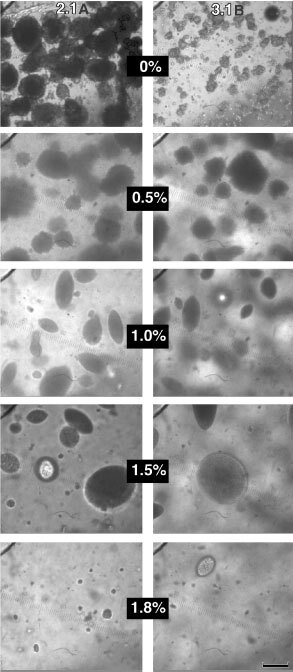
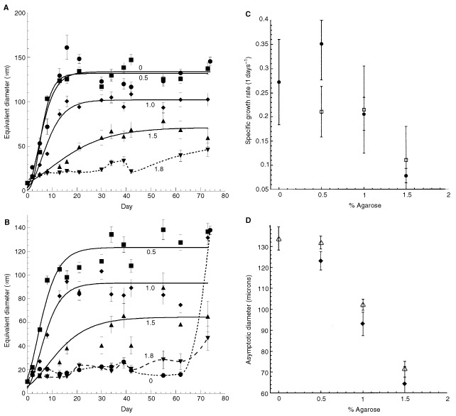
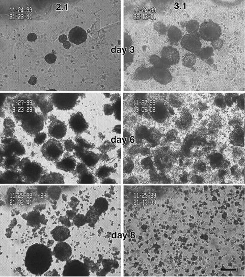
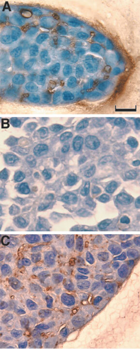
When neoplastic cells grow in confined spaces in vivo, they exert a finite force on the surrounding tissue resulting in the generation of solid stress. By growing multicellular spheroids in agarose gels of defined mechanical properties, we have recently shown that solid stress inhibits the growth of spheroids and that this growth-inhibiting stress ranges from 45 to 120 mmHg. Here we show that solid stress facilitates the formation of spheroids in the highly metastatic Dunning R3327 rat prostate carcinoma AT3.1 cells, which predominantly do not grow as spheroids in free suspension. The maximum size and the growth rate of the resulting spheroids decreased with increasing stress. Relieving solid stress by enzymatic digestion of gels resulted in gradual loss of spheroidal morphology in 8 days. In contrast, the low metastatic variant AT2.1 cells, which grow as spheroids in free suspension as well as in the gels, maintained their spheroidal morphology even after stress removal. Histological examination revealed that most cells in AT2.1 spheroids are in close apposition whereas a regular matrix separates the cells in the AT3.1 gel spheroids. Staining with the hyaluronan binding protein revealed that the matrix between AT3.1 cells in agarose contained hyaluronan, while AT3.1 cells had negligible or no hyaluronan when grown in free suspension. Hyaluronan was found to be present in both free suspensions and agarose gel spheroids of AT2.1. We suggest that cell-cell adhesion may be adequate for spheroid formation, whereas solid stress may be required to form spheroids when cell-matrix adhesion is predominant. These findings have significant implications for tumour growth, invasion and metastasis.
Copyright 2002 Cancer Research UK
Figures

References
-
- BussemakersMJGvan MoorselaarRGorordiLAIchikawaTIsaacsJTTakeshiMDebruyneFMJSchalkenJA1992Decreased expression of E-cadherin in the progression of rat prostatic cancer Cancer Res 5229162922 - PubMed
-
- CarmelietPJainRK2000Angiogenesis in cancer and other diseases Nature 407249257 - PubMed
-
- DowthwaitheGPWardACFlannelyJSuswilloRFLFlanneryCRArcherCWPitsillidesAA1999The effect of mechanical strain on hyaluronan metabolism in embryonic fibrocartilage cells Matrix Biol 18523532 - PubMed
-
- FidlerI1991Cancer Metastasis Br Med Bull 47157177 - PubMed
-
- Griffon-EtienneGBoucherYBrekkenCSuitHDJainRK1999Taxane-induced apoptosis decompresses blood vessels and lowers interstitial fluid pressure in solid tumors: Clinical implications Cancer Res 5937763782 - PubMed
Publication types
MeSH terms
Substances
Grants and funding
LinkOut - more resources
Full Text Sources
Other Literature Sources
Miscellaneous
