. 2002 Apr 22;86(8):1262-9.
doi: 10.1038/sj.bjc.6600264.

Molecular changes in the expression of human colonic nutrient transporters during the transition from normality to malignancy

Affiliations
Free PMC article

Molecular changes in the expression of human colonic nutrient transporters during the transition from normality to malignancy

D W Lambert et al. Br J Cancer. .
Free PMC article

Abstract

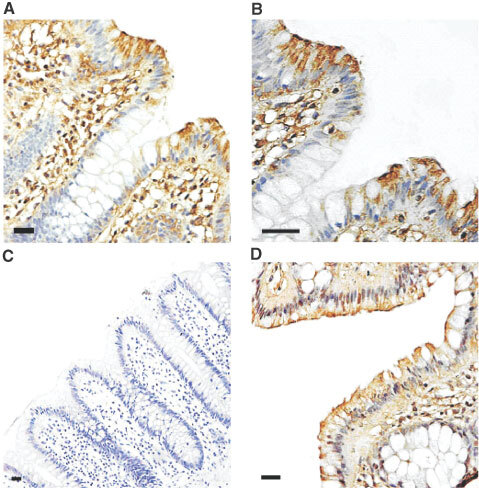
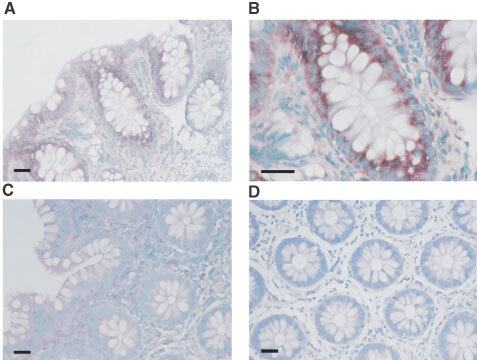
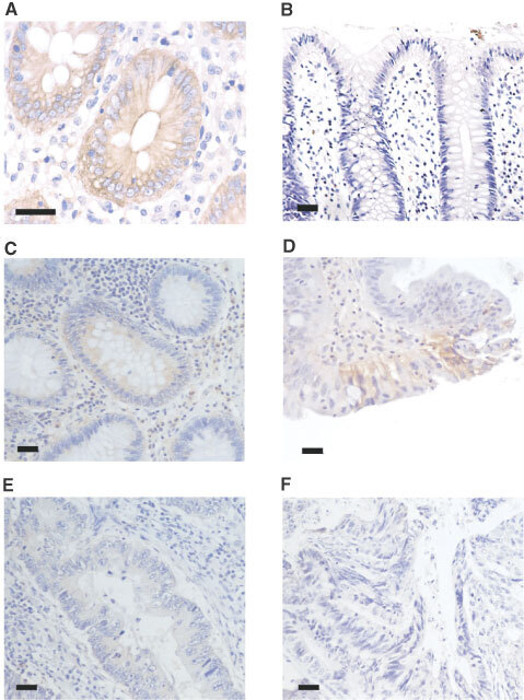
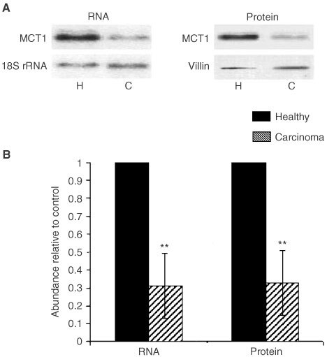
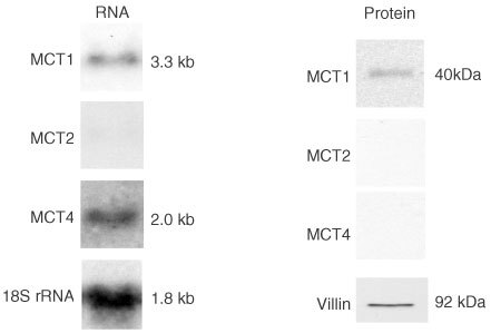
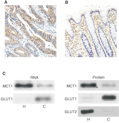
Healthy colonocytes derive 60-70% of their energy supply from short-chain fatty acids, particularly butyrate. Butyrate has profound effects on differentiation, proliferation and apoptosis of colonic epithelial cells by regulating expression of various genes associated with these processes. We have previously shown that butyrate is transported across the luminal membrane of the colonic epithelium via a monocarboxylate transporter, MCT1. In this paper, using immunohistochemistry and in situ hybridisation histochemistry, we have determined the profile of MCT1 protein and mRNA expression along the crypt to surface axis of healthy human colonic tissue. There is a gradient of MCT1 protein expression in the apical membrane of the cells along the crypt-surface axis rising to a peak in the surface epithelial cells. MCT1 mRNA is expressed along the crypt-surface axis and is most abundant in cells lining the crypt. Analysis of healthy colonic tissues and carcinomas using immunohistochemistry and Western blotting revealed a significant decline in the expression of MCT1 protein during transition from normality to malignancy. This was reflected in a corresponding reduction in MCT1 mRNA expression, as measured by Northern analysis. Carcinoma samples displaying reduced levels of MCT1 were found to express the high affinity glucose transporter, GLUT1, suggesting that there is a switch from butyrate to glucose as an energy source in colonic epithelia during transition to malignancy. The expression levels of MCT1 in association with GLUT1 could potentially be used as determinants of the malignant state of colonic tissue.


Figures

Figure 1
Figure 1
Immunohistochemical detection of MCT1 in healthy human colon. Immunohistochemistry for MCT1 and villin was carried out on healthy human colon biopsy sections using a peroxidase-conjugated secondary antibody as described in the Materials and Methods section. MCT1 staining is seen as a brown colouring contrasting with the blue/purple counterstained nuclei. (A) MCT1, ×200 magnification; (B) MCT1, ×400 magnification; (C) MCT1 antibody pre-incubated with immunising peptide, ×100 magnification; (D) immunodetection of villin, ×200 magnification. Bars represent 50 μm.
Figure 2
Figure 2
Detection of MCT1 mRNA in healthy human colonic biopsy sections by in situ hybridisation. In situ hybridisation was carried out on healthy human colon biopsy sections using DIG labelled cRNA probes as described in the Materials and Methods section. Sections were hybridised with a specific antisense probe for MCT1 and the corresponding sense probe to act as a control for non-specific binding. (A) MCT1 antisense probe (×200); (B) MCT1 antisense probe (×400); (C) sense probe (×200); (D) MCT1 antisense probe on RNase pre-treated sections (×200). Specific staining is seen as a dark purple colour, contrasting with the blue/green counterstained nuclei. Bars represent 50 μm.
Figure 3
Figure 3
Immunohistochemical detection of MCT1 in normal, adenoma and carcinoma human colonic sections. Immunohistochemistry was carried out on normal, adenoma and carcinoma human colonic tissue using a horseradish peroxidase-conjugated secondary antibody as described in the Materials and Methods section. MCT1 staining is seen as a brown colouring contrasting with the blue/purple counterstained nuclei. (A) Healthy human colon (×400); (B) healthy human colon with MCT1 antibody pre-incubated with immunising peptide (×200); (C) tubular adenoma (×200); (D) tubular-villous adenoma (×200); (E) well differentiated carcinoma (×200); (F) poorly differentiated carcinoma (×200). Bars represent 50 μm. This data is summarised in Table 1.
Figure 4
Figure 4
Assessment of MCT1 protein and mRNA levels in healthy colon and colonic carcinomas by Western and Northern analyses. Post-nuclear membranes and total RNA were isolated from normal and malignant colon, and analysed for MCT1, villin and 18S rRNA abundance as described in the Methods section. (A) Left panel: representative Northern blot showing abundance of MCT1 in healthy and malignant colon. Nylon membranes were stripped and reprobed for 18S rRNA to confirm equality of loading. Right panel: representative Western blot showing abundance of MCT1 in healthy and malignant colon. Nitrocellulose membranes were stripped and re-probed for villin to confirm equality of loading. (B) Combined densitometric analysis of Northern and Western blots displayed as a histogram. MCT1 abundance in carcinomas (shaded square) is displayed relative to healthy (solid square) where abundance in healthy samples=1. All values are normalised to the corresponding 18S rRNA or villin signals. Error bars represent±s.e.m. of 10 paired samples.
Figure 5
Figure 5
Assessment of the expression of MCT2 and MCT 4 in colon carcinoma measured by Western and Northern analyses. Post-nuclear membranes and total RNA were isolated from malignant colonic tissues, and analysed for MCT1, MCT2, MCT4, villin or 18S rRNA expression as described in the Methods section. Left panel: representative Northern blot showing abundance of MCT1 and MCT4 RNA in malignant colon. Nylon membranes were stripped and re-probed for 18S rRNA to confirm equality of loading. Right panel: representative Western blot showing the levels of MCT1 MCT2, and MCT4 proteins in malignant colon carcinoma tissues. Nitrocellulose membranes were stripped and re-probed for villin to confirm equality of protein loading. Note: specific immunoreactive bands were detected in control samples for MCT2 (in rat liver homogenate) and MCT4 (in rat heart tissue homogenate).
Figure 6
Figure 6
Abundance of GLUT1 in healthy and diseased colon. Immunohistochemistry was carried out on healthy and malignant human colonic tissue sections using a horseradish peroxidase conjugated secondary antibody as described under ‘Methods’. GLUT1 staining is seen as a brown colour contrasting with the blue/purple counterstained nuclei. (A) moderately differentiated carcinoma (×200). Bars represent 50 μm. Pre-incubation of antibody with immunising peptide resulted in absence of staining (data not shown). (B) Healthy colon (×200). Significant erythrocyte staining can be seen in the lamina propria. (C) Post-nuclear membranes and total RNA were extracted from healthy (H) and carcinoma (C) tissues and analysed for MCT1, GLUT1 and GLUT2 mRNA and protein abundance as described in ‘Methods’. Left panel: representative Northern blot indicating levels of MCT1 and GLUT1 mRNA (n=6). Right panel: Representative Western blot demonstrating levels of MCT1, GLUT1 and GLUT2 proteins (n=6).

References

    1. ArcherSYMengSSheiAHodinRA1998p21(WAF1) is required for butyrate-mediated growth inhibition of human colon cancer cells PNAS 9567916796 - PMC - PubMed
    1. BaldwinSA1993Mammalian passive glucose transporters: members of a ubiquitous family of active and passive transport proteins Biochim Biophys Act 11541749 - PubMed
    1. BrownRSWahlRL1993Overexpression of Glut-1 glucose transporter in human breast cancer Cancer 7229792985 - PubMed
    1. BugautMBentejacM1993Biological effects of short-chain fatty acids in nonruminant mammals Ann Rev Nutr 13217241 - PubMed
    1. BurkittDP1971Epidemiology of cancer of the colon and rectum Cancer 28313 - PubMed

Publication types

MeSH terms